

Intraoperative 2D C-arm and 3D O-arm in children: a comparative phantom study
- PMID: 30294382
- PMCID: PMC6169555
- DOI: 10.1302/1863-2548.12.180016
Intraoperative 2D C-arm and 3D O-arm in children: a comparative phantom study
Abstract
Purpose: Exposure to ionizing radiation is a concern for children during intraoperative imaging. We aimed to assess the radiation exposure to the paediatric patient with 2D and 3D imaging.
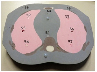
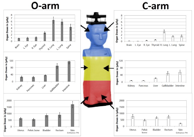
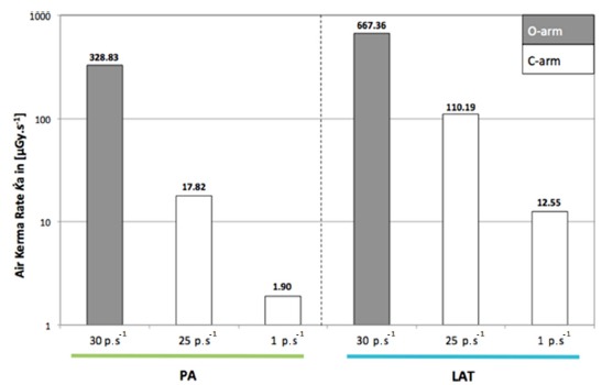
Methods: To evaluate the radiation exposure, patient absorbed doses to the organs were measured in an anthropomorphic phantom representing a five-year-old child, using thermoluminescent dosimeters. For comparative purposes, organ doses were measured using a C-arm for one minute of fluoroscopy and one acquisition with an O-arm. The cone-beam was centred on the pelvis. Direct and scattered irradiations were measured and compared (Student's t-test). Skin entrance dose rates were also evaluated.
Results: All radiation doses were expressed in µGy. Direct radiation doses of pelvic organs were between 631.22 and 1691.87 for the O-arm and between 214.08 and 737.51 for the C-arm, and were not significant (p = 0.07). Close scattered radiation on abdominal organs were between 25.11 and 114.85 for the O-arm and between 8.03 and 55.34 for the C-arm, and were not significant (p = 0.07). Far scattered radiation doses on thorax, neck and head varied from 0.86 to 6.42 for the O-arm and from 0.04 to 3.08 for the C-arm, and were significant (p = 0.02). The dose rate at the skin entrance was 328.58 µGy.s-1 for the O-arm and 1.90 with the C-arm.
Conclusion: During imaging of the pelvis, absorbed doses for a 3D O-arm acquisition were higher than with one minute fluoroscopy with the C-arm. Further clinical studies comparing effective doses are needed to assess ionizing risks of the intraoperative imaging systems in children.
Keywords: CBCT; O-arm; ionizing radiations; paediatric orthopaedics; patient exposure.
Figures
References
-
- National Research Council Health risks from exposure to low levels of ionizing radiation: BEIR VII phase 2. National Academies Press 2006. April 23. - PubMed
-
- Shore RE, Hildreth N, Dvoretsky P, et al. Thyroid cancer among persons given X-ray treatment in infancy for an enlarged thymus gland. Am J Epidemiol 1993;137:1068–1080. - PubMed
-
- Preston DL, Mattsson A, Holmberg E, et al. Radiation effects on breast cancer risk: a pooled analysis of eight cohorts. Radiat Res 2002;158:220–235. - PubMed
-
- Karlsson P, Holmberg E, Lundell M, et al. Intracranial tumors after exposure to ionizing radiation during infancy: a pooled analysis of two Swedish cohorts of 28,008 infants with skin hemangioma. Radiat Res 1998;150:357–364. - PubMed
LinkOut - more resources
Full Text Sources
