

Diagnostic Performance of Deep Learning Algorithms Applied to Three Common Diagnoses in Dermatopathology
- PMID: 30294501
- PMCID: PMC6166480
- DOI: 10.4103/jpi.jpi_31_18
Diagnostic Performance of Deep Learning Algorithms Applied to Three Common Diagnoses in Dermatopathology
Abstract
Background: Artificial intelligence is advancing at an accelerated pace into clinical applications, providing opportunities for increased efficiency, improved accuracy, and cost savings through computer-aided diagnostics. Dermatopathology, with emphasis on pattern recognition, offers a unique opportunity for testing deep learning algorithms.
Aims: This study aims to determine the accuracy of deep learning algorithms to diagnose three common dermatopathology diagnoses.
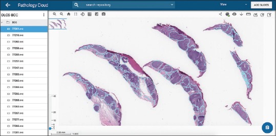
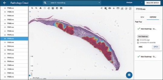
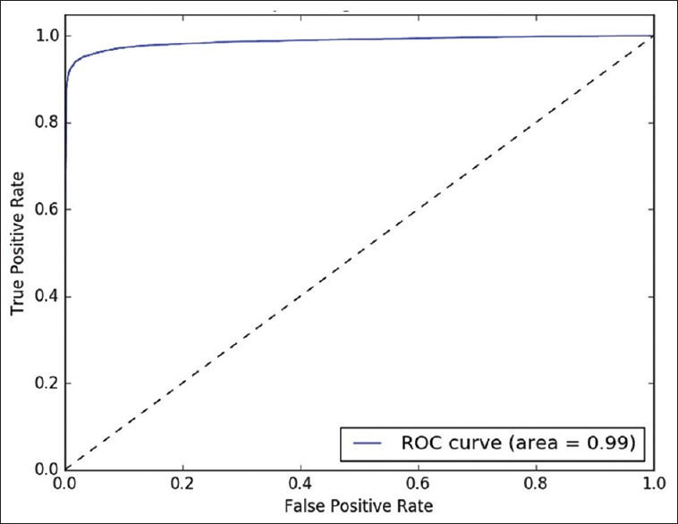
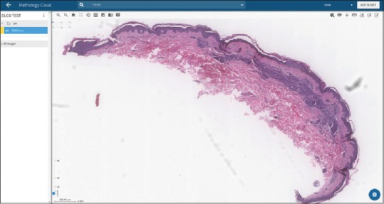
Methods: Whole slide images (WSI) of previously diagnosed nodular basal cell carcinomas (BCCs), dermal nevi, and seborrheic keratoses were annotated for areas of distinct morphology. Unannotated WSIs, consisting of five distractor diagnoses of common neoplastic and inflammatory diagnoses, were included in each training set. A proprietary fully convolutional neural network was developed to train algorithms to classify test images as positive or negative relative to ground truth diagnosis.
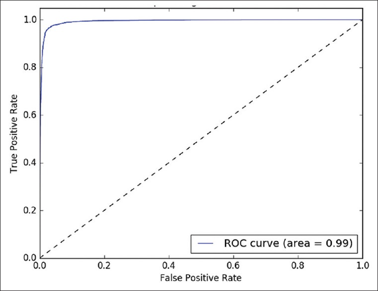
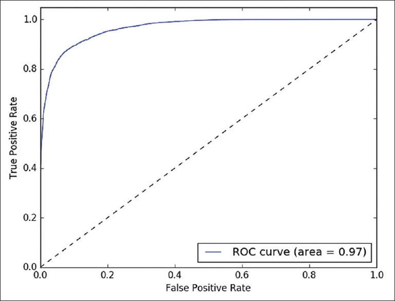
Results: Artificial intelligence system accurately classified 123/124 (99.45%) BCCs (nodular), 113/114 (99.4%) dermal nevi, and 123/123 (100%) seborrheic keratoses.
Conclusions: Artificial intelligence using deep learning algorithms is a potential adjunct to diagnosis and may result in improved workflow efficiencies for dermatopathologists and laboratories.
Keywords: Artificial intelligence; computational pathology; computer-aided diagnosis; deep learning algorithm; dermatopathology; digital pathology; whole slide images.
Conflict of interest statement
Dr. Thomas Olsen is an owner at DLCS-Clearpath, LLC.
Figures
References
-
- Klipp J. Poughkeepsie NY: Laboratory Economics; 2017. The US Anatomic Pathology Market: Forecasts and Trends 2017-2020; pp. 79–80.
-
- Zembowicz A, Ahmad A, Lyle SR. A comprehensive analysis of a web-based dermatopathology second opinion consultation practice. Arch Pathol Lab Med. 2011;135:379–83. - PubMed
-
- Nielsen PS, Riber-Hansen R, Raundahl J, Steiniche T. Automated quantification of MART1-verified Ki67 indices by digital image analysis in melanocytic lesions. Arch Pathol Lab Med. 2012;136:627–34. - PubMed
-
- Al-Janabi S, Huisman A, Van Diest PJ. Digital pathology: Current status and future perspectives. Histopathology. 2012;61:1–9. - PubMed
LinkOut - more resources
Full Text Sources
Other Literature Sources
