

Potentials and challenges of diffusion-weighted magnetic resonance imaging in radiotherapy
- PMID: 30294681
- PMCID: PMC6169338
- DOI: 10.1016/j.ctro.2018.09.002
Potentials and challenges of diffusion-weighted magnetic resonance imaging in radiotherapy
Abstract
Purpose: To review the potential and challenges of integrating diffusion weighted magnetic resonance imaging (DWI) into radiotherapy (RT).
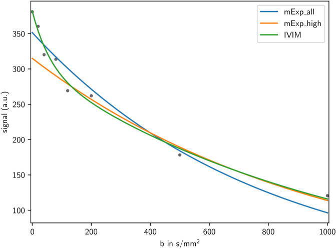
Content: Details related to image acquisition of DWI for RT purposes are discussed, along with the challenges with respect to geometric accuracy and the robustness of quantitative parameter extraction. An overview of diffusion- and perfusion-related parameters derived from mono- and bi-exponential models is provided, and their role as potential RT biomarkers is discussed. Recent studies demonstrating potential of DWI in different tumor sites such as the head and neck, rectum, cervix, prostate, and brain, are reviewed in detail.
Conclusion: DWI has shown promise for RT outcome prediction, response assessment, as well as for tumor delineation and characterization in several cancer types. Geometric and quantification robustness is challenging and has to be addressed adequately. Evaluation in larger clinical trials with well designed imaging protocol and advanced analysis models is needed to develop the optimal strategy for integrating DWI in RT.
Figures
References
-
- Sander L., Langkilde N.C., Holmberg M., Carl J. MRI target delineation may reduce long-term toxicity after prostate radiotherapy. Acta Oncol. 2014;53(6):809–814. - PubMed
-
- Thorwarth D., Leibfarth S., Mönnich D. Potential role of PET/MRI in radiotherapy treatment planning. Clin Transl Imaging. 2013;1(1):45–51.
Publication types
LinkOut - more resources
Full Text Sources
