. 2018 Dec;84(6):854-872.
doi: 10.1002/ana.25356. Epub 2018 Nov 29.

N-acetylcysteine targets 5 lipoxygenase-derived, toxic lipids and can synergize with prostaglandin E2 to inhibit ferroptosis and improve outcomes following hemorrhagic stroke in mice

Affiliations

N-acetylcysteine targets 5 lipoxygenase-derived, toxic lipids and can synergize with prostaglandin E2 to inhibit ferroptosis and improve outcomes following hemorrhagic stroke in mice

Saravanan S Karuppagounder et al. Ann Neurol. 2018 Dec.

Abstract

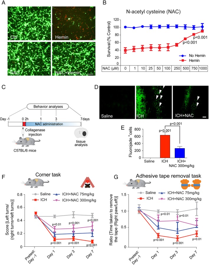
Objectives: N-acetylcysteine (NAC) is a clinically approved thiol-containing redox modulatory compound currently in trials for many neurological and psychiatric disorders. Although generically labeled as an "antioxidant," poor understanding of its site(s) of action is a barrier to its use in neurological practice. Here, we examined the efficacy and mechanism of action of NAC in rodent models of hemorrhagic stroke.

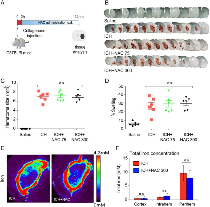
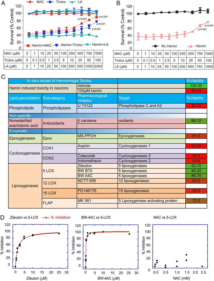
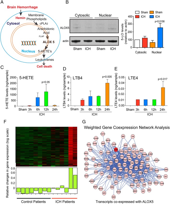
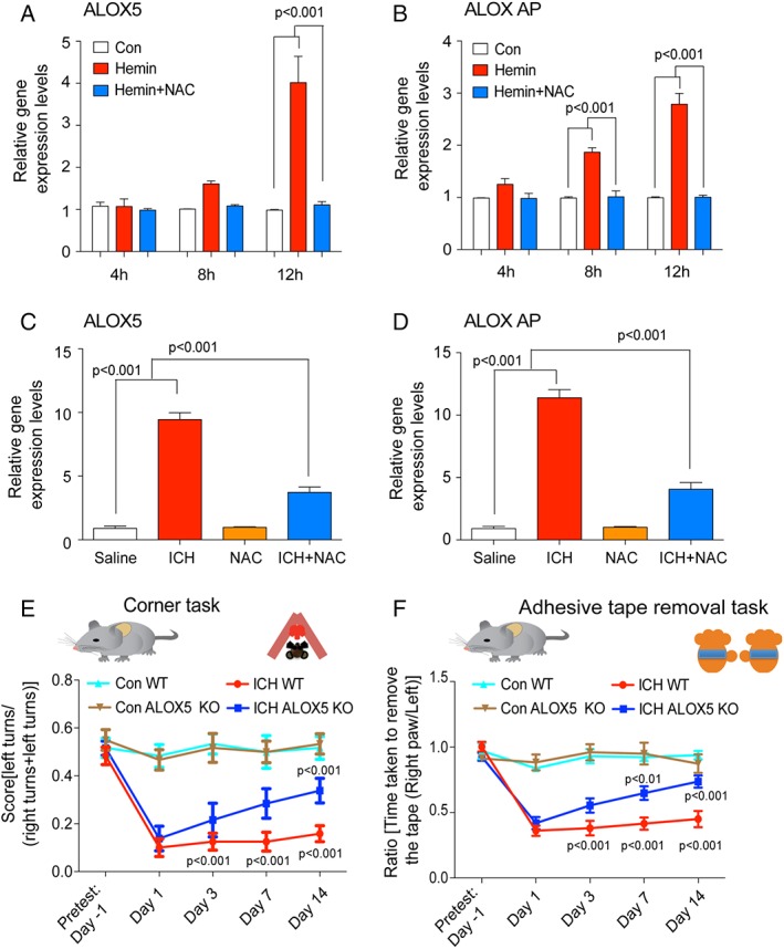
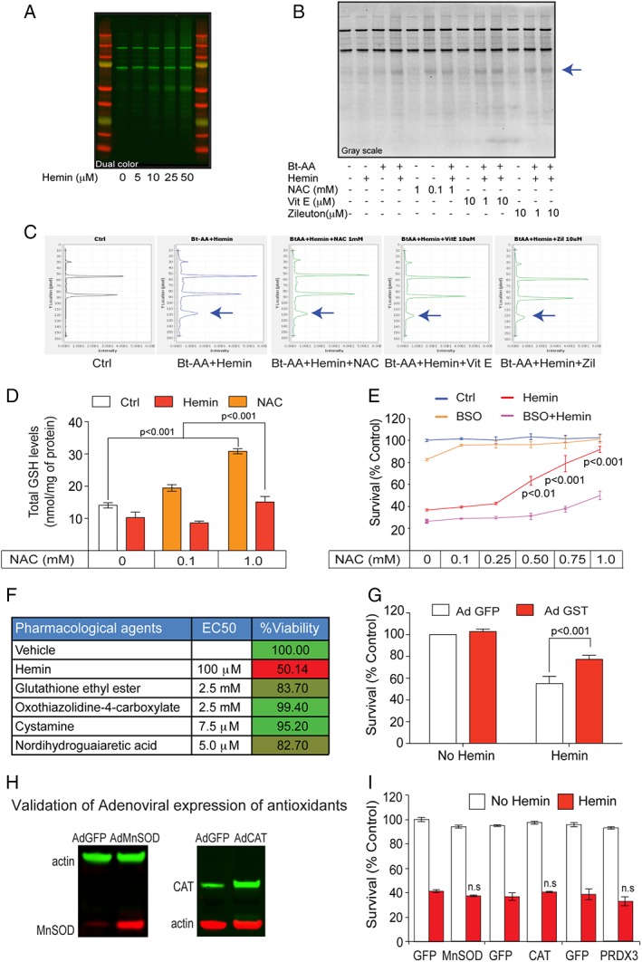
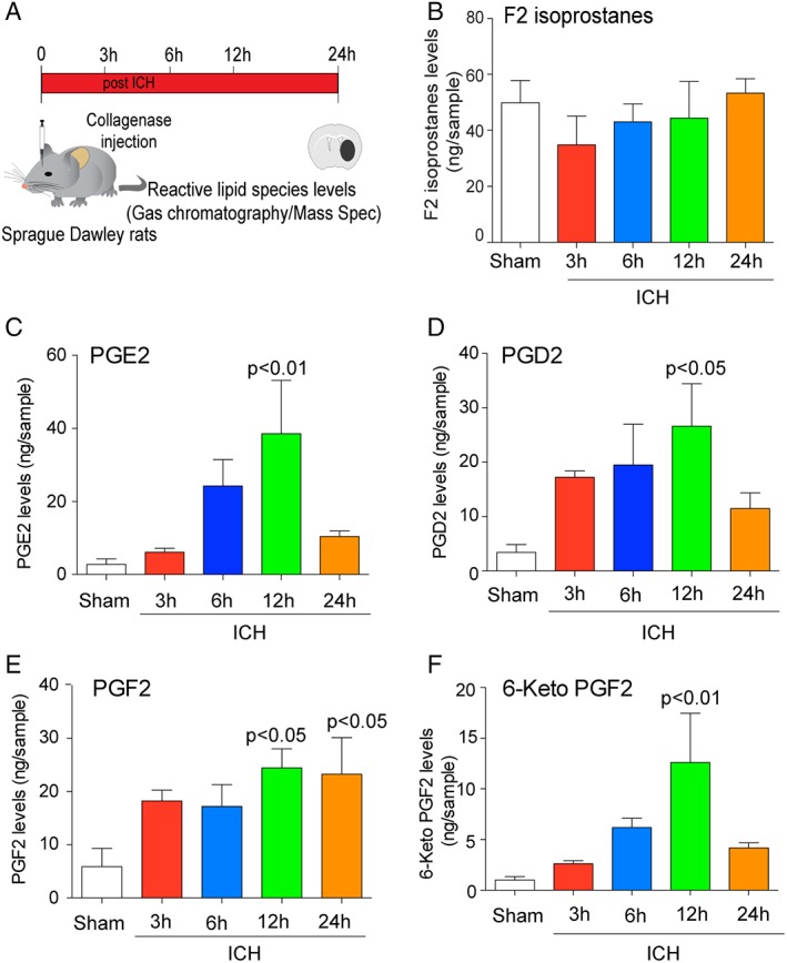
Methods: Hemin was used to model ferroptosis and hemorrhagic stroke in cultured neurons. Striatal infusion of collagenase was used to model intracerebral hemorrhage (ICH) in mice and rats. Chemical biology, targeted lipidomics, arachidonate 5-lipoxygenase (ALOX5) knockout mice, and viral-gene transfer were used to gain insight into the pharmacological targets and mechanism of action of NAC.

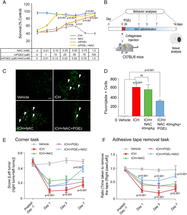
Results: NAC prevented hemin-induced ferroptosis by neutralizing toxic lipids generated by arachidonate-dependent ALOX5 activity. NAC efficacy required increases in glutathione and is correlated with suppression of reactive lipids by glutathione-dependent enzymes such as glutathione S-transferase. Accordingly, its protective effects were mimicked by chemical or molecular lipid peroxidation inhibitors. NAC delivered postinjury reduced neuronal death and improved functional recovery at least 7 days following ICH in mice and can synergize with clinically approved prostaglandin E2 (PGE2 ).

Interpretation: NAC is a promising, protective therapy for ICH, which acted to inhibit toxic arachidonic acid products of nuclear ALOX5 that synergized with exogenously delivered protective PGE2 in vitro and in vivo. The findings provide novel insight into a target for NAC, beyond the generic characterization as an antioxidant, resulting in neuroprotection and offer a feasible combinatorial strategy to optimize efficacy and safety in dosing of NAC for treatment of neurological disorders involving ferroptosis such as ICH. Ann Neurol 2018;84:854-872.


Conflict of interest statement

R.R.R. and S.S.K. are co‐inventors on a patent related to the use of N‐acetylcysteine and/or PGE2 in neurological disorders. These patents have been licensed by Neuronasal, Inc., an early‐stage biotechnology company. R.R.R. is on the SAB for Neuronasal and has a small equity interest in the company along with receiving occasional consulting fees.

Figures

Figure 1
Figure 1
N‐acetylcysteine (NAC) abrogated hemin‐induced ferroptosis in primary neurons in vitro and reduced cell death and enhanced functional recovery in a collagenase ICH model in mice. (A) Representative images of primary cortical neurons 24 hours after treatment with saline, NAC (1mM), hemin (100 μM) and hemin (100 μM) + NAC (1mM). LIVE/DEAD assay; green fluorescent cells labeled with calcein‐AM are alive; red fluorescent cells labeled with ethidium homodimer are dead. Scale bars, 100 μm. (B) NAC protected primary cortical neurons from hemin‐induced ferroptosis in a concentration‐dependent manner. Cell death was analyzed 24 hours after hemin treatment with or without NAC by monitoring MTT reduction, a population measure of cell viability. (C) Experimental design for delivery of NAC post‐ICH in mice. NAC (75mg and 300mg/kg; intraperitoneal) was delivered 2 hours after collagenase injection and then daily for 7 days. Behavior was assessed using the corner task (spatial neglect) and adhesive tape removal task (sensory neglect) and was assessed on days 1, 3, and 7 after ICH. (D) NAC reduced neuronal degeneration was monitored by Fluoro‐Jade (FJ) staining (green) in the perihematomal regions of the mouse brain. Representative images show increased numbers of degenerating neurons (white arrows) in the ICH‐treated group; this was reduced by NAC treatment. Scale bar, 100 μm. (E) Quantification of FJ staining of neurons, a nonspecific marker of degeneration, after NAC treatment in ICH mice brains. (F) NAC (300mg/kg) significantly reduces spatial neglect associated with ICH. (G) NAC (300mg/kg) reduces sensory neglect (adhesive tape removal task) induced by ICH (n = 11). NAC (75mg/kg) had no significant effect in mice or rats (not shown). Significance was determined by two‐way ANOVA and Bonferroni's post‐hoc test. All graphs are mean ± SEM. ANOVA = analysis of variance; ICH = intracerebral hemorrhage; NAC = N‐acetylcysteine; MTT = methyl thiazolyl tetrazolium; SEM = standard error of the mean.
Figure 2
Figure 2
N‐acetylcysteine (NAC) enhanced functional recovery following intracerebral hemorrhage (ICH) without influencing collagenase activity in vivo or iron levels or iron distribution in the brain after ICH. (A) Schematic illustration of NAC treatment, post‐ICH. (B) Serial brain sections from saline‐, ICH‐, and ICH + NAC–treated groups. (C,D) Quantification of hematoma size and brain edema by light microscopy revealed no significant difference between control and NAC‐treated groups in mice (n = 6). (E) x‐ray fluorescence microscopy analysis of pseudo‐colored images from coronal sections of collagenase alone or NAC treatment in collagenase‐induced ICH in mice after 7 days. Quantification in cortex, hematomal, and perihematomal regions revealed that total iron does not change in the brain following NAC treatment (F). Significance determined by one‐way ANOVA followed by Dunnett's comparison test, for vehicle treatment ICH or NAC treatment (C,D). All graphs are mean ± SEM. ANOVA = analysis of variance; ICH = intracerebral hemorrhage; NAC = N‐acetylcysteine; ns = not significant; SEM = standard error of the mean.
Figure 3
Figure 3
Systematic pharmacological characterization of arachidonic acid metabolizing enzymes in hemin‐induced ferroptosis in primary cortical neurons identified 5‐Lipoxygenase (ALOX5) as a target. (A) N‐acetylcysteine (NAC), a cysteine prodrug, or Trolox (TRO) dose dependently prevented hemin‐induced ferroptosis in primary cortical neurons as measured by MTT assay. Lipoic acid (LA) failed to prevent hemin‐induced ferroptosis in primary cortical neurons. (B) Combinations of nonprotective concentrations of NAC, TRO, or LA failed to synergize in preventing hemin‐induced ferroptosis in primary cortical neurons. (C) Table illustrates that structurally diverse inhibitors of 5‐lipoxygenase, but not inhibitors of other arachidonate metabolizing enzymes, prevent hemin‐induced ferroptosis in primary cortical neurons. (D) Assay of recombinant ALOX5 activity in vitro (test tube) showed that putative ALOX5 inhibitors are potently effective in inhibiting the enzyme, whereas NAC is not. Significance was determined by two‐way ANOVA and Bonferroni's post‐hoc test. ANOVA = analysis of variance; MTT = methyl thiazolyl tetrazolium.
Figure 4
Figure 4
Intracerebral hemorrhage (ICH)‐induced ALOX5‐derived oxidized lipids and gene expression in mice and humans. (A) Schematic model of ALOX5 pathway activation in ICH. (B) ICH increased ALOX5 protein levels in the nuclear fraction as verified by immunoblot analysis. Gas chromatography/mass spectrometry analysis revealed a significant increase in ALOX5‐derived lipid species after ICH in rats (n = 4) compared to sham, including 5‐hydroxyeicosatetraenoic acid (5‐HETE) (C), Leukotriene B4 (LTB4) (D), and Leukotriene E4 (E). Data from sham control brains from each time point were pooled for the analysis. (F) Transcriptomic analysis of brain tissues from control (n = 8) and ICH (n = 6) patients. (G) Weighted gene coexpression network analysis (WGCNA) revealed transcripts most closely coregulated with ALOX5. Significance was determined by one‐way ANOVA and Dunnet's multiple‐comparison test. All graphs are mean ± SEM. ANOVA = analysis of variance; ICH = intracerebral hemorrhage; SEM = standard error of the mean.
Figure 5
Figure 5
N‐acetylcysteine (NAC) reduced ICH‐induced ALOX5 gene expression and molecular knockdown of ALOX5 improves ICH‐induced behavioral deficits. RT‐PCR analysis revealed that toxic levels of hemin time dependently (4, 8, and 12 hours) increased ALOX5 and ALOX activating protein (AP) levels in primary neurons. Protective doses of NAC (1mM) blocked this expression in primary cortical neurons (A,B). In mice, NAC significantly blocked ICH‐induced ALOX5 (C) and ALOX AP levels (D). Molecular knockdown of ALOX5 improved spatial neglect behavioral deficits and sensory neglect (adhesive tape removal task) behavioral deficits associated with ICH compared to WT controls. Significance was determined by two‐way ANOVA and Bonferroni's post‐hoc test. All graphs are mean ± SEM. ANOVA = analysis of variance; ICH = intracerebral hemorrhage; NAC = N‐acetylcysteine; SEM = standard error of the mean; WT, wild type.
Figure 6
Figure 6
Protective NAC reduced lipid protein adducts and required glutathione (GSH) synthesis for neuroprotection. (A) Primary cortical neurons were treated with progressively higher concentrations of hemin. Lipid protein adducts (arachidonic acid‐biotin) were detected by western blots probed with streptavidin‐HRP (horseradish peroxidase). (B) NAC (1mM), α‐tocopherol (10 μM), and Zileuton (10 μM) attenuated hemin‐induced oxidized lipid protein adducts. Grayscale image of lipid protein adducts probed with streptavidin‐HRP. Representative blot from replicates of three experiments. (C) Quantification of bands reveals a significant reduction in oxidized lipid protein adducts after NAC, Zileuton, and vitamin E. (D) High‐performance liquid chromatography analysis of total GSH revealed that NAC increases GSH levels in control and hemin‐treated cortical neurons. (E) Pharmacological inhibition of γ‐glutamylcysteine synthetase, the rate‐limiting enzyme in GSH synthesis, by BSO blocked the ability of NAC to prevent hemin‐induced ferroptosis in primary cortical neurons. (F) As predicted from results in (B), table shows that multiple established strategies to increase GSH levels in neurons or glia prevented hemin‐induced ferroptosis. GSH ethyl ester (a membrane permeant form of GSH); L‐oxothiazolidine‐4‐carboxylate (OTC), a cysteine donor; Nrf2 activators cystamine and NDGA, which increase GSH synthesizing and GSH‐dependent detoxification enzymes, abrogate hemin‐induced ferroptosis in primary cortical neurons as measured by MTT assay. (G) Adenoviral overexpression of GSTA4, a GSH enzyme known to neutralize toxic lipids, protects against hemin‐induced ferroptosis. (H) Representative immunoblot for forced expression of antioxidants. (I) Overexpression of MnSOD, catalase, and peroxiredoxin 3 failed to protect against hemin‐induced toxicity. Significance was determined by two‐way ANOVA followed by Bonferroni's comparison test. All graphs are mean ± SEM. Ad GFP = adenoviral green fluorescent protein; ANOVA = analysis of variance; BSO = buthionine sulfoximine; EC50 = half maximal effective concentration; MTT = methyl thiazolyl tetrazolium; NAC = N‐acetylcysteine; ns = not significant; SEM = standard error of the mean.
Figure 7
Figure 7
Intracerebral hemorrhage (ICH) in rodents induced COX‐derived oxidized lipids. (A) Experimental design for analyzing eicosanoid levels post‐ICH in mice. (B) GC/MS analysis revealed that levels of F2 isoprostanes, established markers of nonenzymatic lipid peroxidation, were not altered after ICH (n = 4). COX‐derived species PGE2 (C), PGD2 (D), PGF2 (E), and 6‐keto PGF2 (F) were time dependently increased post‐ICH (n = 4). Data from sham control brains from each time points were pooled for the analysis. Significance was determined by one‐way ANOVA and Dunnet's multiple‐comparison test. All graphs are mean ± SEM. ANOVA = analysis of variance; COX = cyclooxygenase; GC/MS = gas chromatography/mass spectrometry; ICH = intracerebral hemorrhage; PGD2 = prostaglandin D2; PGE2 = prostaglandin E2; PGF2 = prostaglandin F2; SEM = standard error of the mean.
Figure 8
Figure 8
Prostaglandin (PGE2) synergized with NAC to prevent hemin‐induced ferroptosis in vitro and in improving functional recovery following ICH in mice in vivo. (A) NAC in combination with PGE2 synergized against hemin‐induced ferroptosis in primary cortical neurons. (B) Schematic illustration of the combinatorial delivery of NAC (40mg/kg, intraperitoneal) and PGE2 (10 μM, intracerebroventricular) after ICH in mice. The corner task (spatial neglect) and adhesive tape removal task (sensory neglect) were assessed on days 1, 3, and 7 after ICH to assess behavioral improvement. (C) The NAC (40 mg/kg, intraperitoneal) and intracerebroventricular (10 μM) PGE2 combination reduced neuronal degeneration as monitored by Fluoro‐Jade staining (green) in the perihematomal regions of the mouse brain after ICH. (D,E) Intraperitoneal NAC (40mg/kg) plus intracerebroventricular PGE2 reduces spatial neglect and sensory neglect induced by ICH, whereas each agent alone does not (n = 10). Significance was determined by two‐way ANOVA and Bonferroni's post hoc test. All graphs are mean ± SEM. ANOVA = analysis of variance; ICH = intracerebral hemorrhage; NAC = N‐acetylcysteine; ns = not significant; PGE2 = prostaglandin E2; SEM = standard error of the mean.

Similar articles

Cited by

References

    1. Qureshi AI, Suri MF, Georgiadis AL, et al. Intra‐arterial recanalization techniques for patients 80 years or older with acute ischemic stroke: pooled analysis from 4 prospective studies. AJNR Am J Neuroradiol 2009;30:1184–1189. - PMC - PubMed
    1. van Asch CJ, Oudendijk JF, Rinkel GJ, Klijn CJ. Early intracerebral hematoma expansion after aneurysmal rupture. Stroke 2010;41:2592–2595. - PubMed
    1. Green JL, Heard KJ, Reynolds KM, Albert D. Oral and intravenous acetylcysteine for treatment of acetaminophen toxicity: a systematic review and meta‐analysis. West J Emerg Med 2013;14:218–226. - PMC - PubMed
    1. Dekhuijzen PN, van Beurden WJ. The role for N‐acetylcysteine in the management of COPD. Int J Chron Obstruct Pulmon Dis 2006;1:99–106. - PMC - PubMed
    1. De Rosa SC, Zaretsky MD, Dubs JG, et al. N‐acetylcysteine replenishes glutathione in HIV infection. Eur J Clin Invest 2000;30:915–929. - PubMed

Publication types

MeSH terms