Comparative Study
. 2019 Mar;16(2):160-170.
doi: 10.1177/1479164118802550. Epub 2018 Oct 8.

Effects of obesity on insulin: insulin-like growth factor 1 hybrid receptor expression and Akt phosphorylation in conduit and resistance arteries

Affiliations
Comparative Study

Effects of obesity on insulin: insulin-like growth factor 1 hybrid receptor expression and Akt phosphorylation in conduit and resistance arteries

Romana S Mughal et al. Diab Vasc Dis Res. 2019 Mar.

Abstract

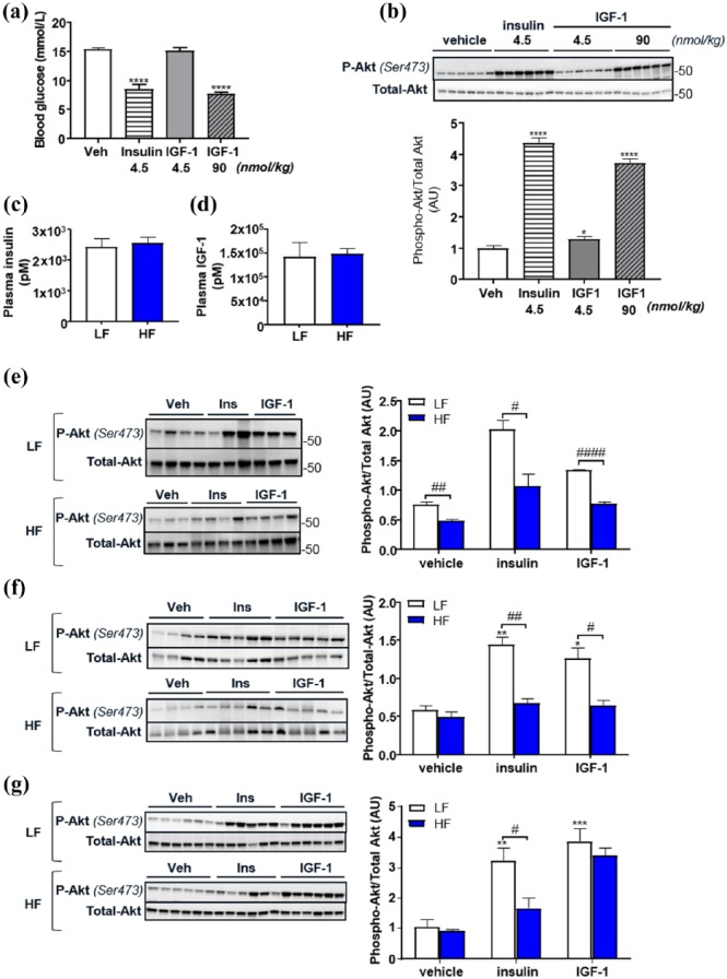
Insulin and insulin-like growth factor-1 stimulate specific responses in arteries, which may be disrupted by diet-induced obesity. We examined (1) temporal effects of high-fat diet compared to low-fat diet in mice on insulin receptor, insulin-like growth factor-1 receptor, insulin receptor/insulin-like growth factor-1 receptor hybrid receptor expression and insulin/insulin-like growth factor-1-mediated Akt phosphorylation in aorta; and (2) effects of high-fat diet on insulin and insulin-like growth factor-1-mediated Akt phosphorylation and vascular tone in resistance arteries. Medium-term high-fat diet (5 weeks) decreased insulin-like growth factor-1 receptor expression and increased hybrid expression (~30%) only. After long-term (16 weeks) high-fat diet, insulin receptor expression was reduced by ~30%, insulin-like growth factor-1 receptor expression decreased a further ~40% and hybrid expression increased a further ~60%. Independent correlates of hybrid receptor expression were high-fat diet, duration of high-fat diet and plasma insulin-like growth factor-1 (all p < 0.05). In aorta, insulin was a more potent activator of Akt than insulin-like growth factor-1, whereas in resistance arteries, insulin-like growth factor-1 was more potent than insulin. High-fat diet blunted insulin-mediated vasorelaxation ( p < 0.01) but had no effect on insulin-like growth factor-1-mediated vasorelaxation in resistance arteries. Our findings support the possibility that hybrid receptor level is influenced by nutritional and metabolic cues. Moreover, vessel-dependent effects of insulin and insulin-like growth factor-1 on vascular tone and Akt activation may have implications in treating obesity-related vascular disease.

Keywords: IGF-1 receptors; Obesity; hybrid receptors.


Conflict of interest statement

Declaration of conflicting interests: The author(s) declared no potential conflicts of interest with respect to the research, authorship and/or publication of this article.

Figures

Figure 1.
Figure 1.
Temporal effects of obesity on IR, IGF-1R and hybrid receptor expression in aorta of low-fat (LF) and high-fat (HF) diet fed mice. Data show changes in (a) IR, (b) IGF-1R and (c) hybrid receptor protein at 2, 5 and 16 weeks of feeding. Representative Western blot images are shown with densitometry (a, b and c, n = 10–25 in each group). Relative (d) IR and (e) IGF-1R mRNA is shown in LF and HF mouse aortae at 16 weeks feeding (n = 6 in each group). Analysis was performed between gels, and samples were normalised to a single control whole cell lysate which was loaded on all gels. All data are given as mean values ± SEM. **p < 0.01, ***p < 0.001, ****p < 0.0001 versus lean group.
Figure 2.
Figure 2.
Effects of supplementation of obesity-related modulators on receptor expression in human umbilical vein endothelial cells in vitro. Data show effects of physiological modulators on (a) IR, (b) IGF-1R and (c) hybrid receptor protein expression. Representative Western blot images are shown with densitometry. All data are given as mean values ± SEM. a: basal (0.5% low-serum medium); b: insulin (100 nM); c: TNF-α (10 ng/mL); d: angiotensin 2 (1 µM); e: H2O2 (50 µM); f: glucose (25 mM); g: glucose (25 mM) + insulin (10 nM); h: IGF-1 (100 nM). *p < 0.05, **p < 0.01 versus control (basal) group (n  = 6 for each).
Figure 3.
Figure 3.
Temporal effects of high-fat (HF) diet-induced obesity on insulin and IGF-1 stimulated Akt phosphorylation compared to lean low-fat (LF) diet fed mice. Reduction in blood glucose is shown in response to insulin (4.5 nmol/kg) and IGF-1 at equimolar (4.5 nmol/kg) and equipotent (90 nmol/kg) doses in lean mice (a). Phosphorylation of Akt in the aorta of lean mice in response to insulin (4.5 nmol/kg) and equipotent and equimolar doses of IGF-1 (b). Data show level of subcutaneously injected human insulin (c) and IGF-1 (d) in plasma of LF and HF diet fed mice. Differences in insulin (4.5 nmol/kg) and IGF-1 (90 nmol/kg) stimulated phosphorylation of Akt in LF and HF mouse aortae are shown at 2 weeks (e), 5 weeks (f) and 16 weeks (g) of feeding. Representative Western blots and densitometry are shown. All data are given as mean values ± SEM (n = 6–8 for each group). Bars represent comparisons made between HF and lean groups. *p < 0.05, **p < 0.01, ***p < 0.001, ****p < 0.0001 versus lean vehicle group.
Figure 4.
Figure 4.
Effects of high fat (HF) diet–induced obesity on mesenteric artery function in response to insulin and IGF-1 compared to lean low fat (LF) diet fed mice. (a) Data show a representative recording of dose-dependent insulin-induced vasorelaxation followed by a time-matched control showing stability of pre-constriction over time. Data show (b) insulin-induced and (c) IGF-1-induced relaxation in pre-constricted mesenteric arteries (first order) taken from LF and HF mice after 16 weeks feeding. Differences in vascular sensitivity to insulin and IGF-1 in (d) LF and (e) HF mice are shown and maximal relaxation achieved with (f) insulin and (g) IGF-1. (h) Insulin and (i) IGF-1-mediated phosphorylation of Akt in LF and HF mesenteric arteries is shown with maximal phosphorylation shown in (i) and (k). All data are given as mean values ± SEM. *p < 0.05, HF versus LF group (n = 3–7 for each group).

References

    1. Tan KT, Luo SC, Ho WZ, et al. Insulin/IGF-1 receptor signalling enhances biosynthetic activity and fat mobilization in the initial phase of starvation in adult male C. elegans. Cell Metab 2011; 14: 390–402. - PubMed
    1. Imrie H, Abbas A, Viswambharan H, et al. Vascular insulin-like growth factor-1 resistance and diet-induced obesity. Endocrinology 2009; 150: 4575–4582. - PubMed
    1. Lawrence MC, McKern NM, Ward CW. Insulin receptor structure and its implications for the IGF-1 receptor. Curr Opin Struct Biol 2007; 17: 699–705. - PubMed
    1. Belfiore A, Frasca F, Pandini G, et al. Insulin receptor isoforms and insulin receptor/insulin-like growth factor receptor hybrids in physiology and disease. Endocr Rev 2009; 30: 586–623. - PubMed
    1. Bailyes EM, Navé BT, Soos MA, et al. Insulin receptor/IGF-I receptor hybrids are widely distributed in mammalian tissues: quantification of individual receptor species by selective immunoprecipitation and immunoblotting. Biochem J 1997; 327: 209–215. - PMC - PubMed

Publication types

MeSH terms