. 2019 Jan 18:690:23-28.
doi: 10.1016/j.neulet.2018.10.004. Epub 2018 Oct 6.

Aging alters glucose uptake in the naïve and injured rodent spinal cord

Affiliations

Aging alters glucose uptake in the naïve and injured rodent spinal cord

Ramona E von Leden et al. Neurosci Lett. .

Abstract

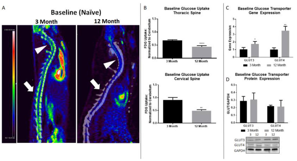
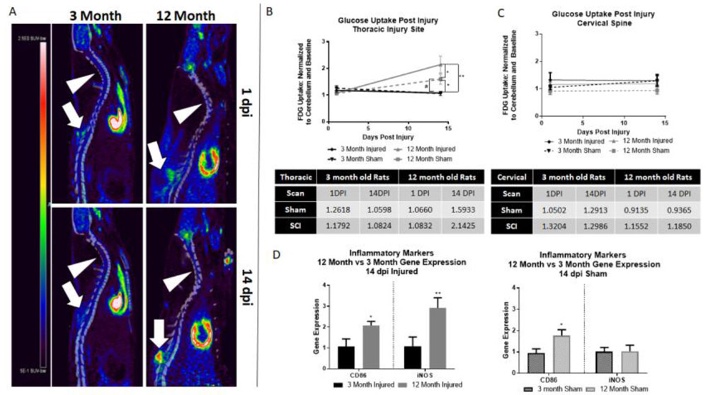
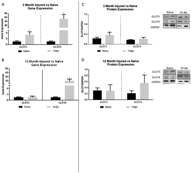
Aging results in increased activation of inflammatory glial cells and decreased neuronal viability following spinal cord injury (SCI). Metabolism and transport of glucose is also decreased with age, although the influence of age on glucose transporter (GLUT) expression or glucose uptake in SCI is currently unknown. We therefore performed [18F]Fluorodeoxyglucose (FDG) PET imaging of young (3 month) and middle-aged (12 month) rats. Glucose uptake in middle-aged rats was decreased compared to young rats at baseline, followed by increased uptake 14 days post contusion SCI. qRT-PCR and protein analysis revealed an association between 14 day glucose uptake and 14 day post-injury inflammation. Further, gene expression analysis of neuron-specific GLUT3 and non-specific GLUT4 (present on glial cells) revealed an inverse relationship between GLUT3/4 gene expression and glucose uptake patterns. Protein expression revealed increased GLUT3 in 3 month rats only, consistent with age related decreases in glucose uptake, and increased GLUT4 in 12 month rats only, consistent with age related increases in inflammatory activity and glucose uptake. Inconsistencies between gene and protein suggest an influence of age-related impairment of translation and/or protein degradation. Overall, our findings show that age alters glucose uptake and GLUT3/4 expression profiles before and after SCI, which may be dependent on level of inflammatory response, and may suggest a therapeutic avenue in addressing glucose uptake in the aging population.

Keywords: Aging; FDG-PET imaging; Glucose metabolism; Glucose transporter; Inflammation; Spinal cord injury.


Figures

Figure 1.
Figure 1.
12 month rats show altered glucose uptake pattern and GLUT3/4 gene expression compared to 3 month rats. A) Representative PET images of 3 and 12 month baseline (naïve) spinal cord. Arrowheads point to cervical spine region, block arrows to thoracic injury site. Heatmap: black/blue represents lowest level of uptake, red/white represents highest level of uptake. B) Naïve 12 month rats show significant decreases in glucose uptake compared to naïve 3 month rats in both the thoracic and cervical cord. No difference in glucose uptake is found in cervical spine after injury, indicating post injury alterations in glucose uptake are specific to the injury site. C) Comparative RT-PCR between 3 month and 12 month rats reveal significantly increased gene expression in 12 month rats in both GLUT3 and GLUT4. D) No significant difference in GLUT3 or GLUT4 protein expression is found by age group. N=8-12/group (PET imaging), 4/group (gene and protein expression); *p<0.05, ** p<0.005, **** p<0.0001.
Figure 2.
Figure 2.
12 month rats show increased glucose uptake after SCI compared to 3 month rats. A) Representative images of both 3 and 12 month rats at 1 and 14 dpi. Arrowheads point to cervical spine, block arrows point to thoracic injury. Heatmap provided to left of images – red represents highest level of uptake. B) 12 month injured rats showed significantly higher glucose uptake than all other groups. 12 month sham rats also show significant increases in glucose uptake compared to 3 month injured (*) and sham (#) groups. Quantitative glucose uptake values in provided table (standard uptake value normalized to cerebellum control and baseline). C) At the cervical spine, no significant difference is found in any group by age or injury. D) When compared to 3 month rats, 12 month injured rats show significantly increased gene expression of both CD86 and iNOS at 14 dpi. 12 month sham rats showed significantly higher CD86 gene expression than 3 month sham rats; no significant difference was seen in iNOS by age group. N=4-6/group; *p<0.05, **p<0.005.
Figure 3.
Figure 3.
Comparative RT-PCR and protein expression for GLUT3 and 4 in 3 and 12 month rats show alterations after SCI. A) 3 month rats show significantly increased GLUT3 and GLUT4 gene expression at 14dpi. B) 12 month rats also show significantly increased GLUT4 at 14 dpi. C) In 3 month rats, GLUT3 protein expression is significantly increased at 14dpi. D) In 12 month rats, GLUT4 protein expression is significantly increased at 14 dpi. Representative images of western blots are shown. N=4/group; *p<0.05.

References

    1. Jain NB, Ayers GD, Peterson EN, Harris MB, Morse L, O’Connor KC, Garshick E, Traumatic spinal cord injury in the United States, 1993–2012, JAMA. 313 (2015) 2236–2243. doi: 10.1001/jama.2015.6250. - DOI - PMC - PubMed
    1. NSCISC, National Spinal Cord Injury Statistical Center, Facts and Figures at a Glance. Birmingham, AL: University of Alabama at Birmingham, 2016, 2016.
    1. Hooshmand MJ, Galvan MD, Partida E, Anderson AJ, Characterization of recovery, repair, and inflammatory processes following contusion spinal cord injury in old female rats: is age a limitation?, Immun. Ageing A 11 (2014) 15. doi: 10.1186/1742-4933-11-15. - DOI - PMC - PubMed
    1. Norden DM, Muccigrosso MM, Godbout JP, Microglial priming and enhanced reactivity to secondary insult in aging, and traumatic CNS injury, and neurodegenerative disease, Neuropharmacology. 96 (2015) 29–41. doi: 10.1016/j.neuropharm.2014.10.028. - DOI - PMC - PubMed
    1. Zhang B, Bailey WM, McVicar AL, Gensel JC, Age increases reactive oxygen species production in macrophages and potentiates oxidative damage after spinal cord injury, Neurobiol. Aging 47 (2016) 157–167. doi: 10.1016/j.neurobiolaging.2016.07.029. - DOI - PMC - PubMed

Publication types