

Macular spatial distribution of preserved autofluorescence in patients with choroideremia
- PMID: 30297337
- PMCID: PMC6533159
- DOI: 10.1136/bjophthalmol-2018-312620
Macular spatial distribution of preserved autofluorescence in patients with choroideremia
Abstract
Background/aims: To better understand the pattern of degeneration progression in cases with choroideremia.
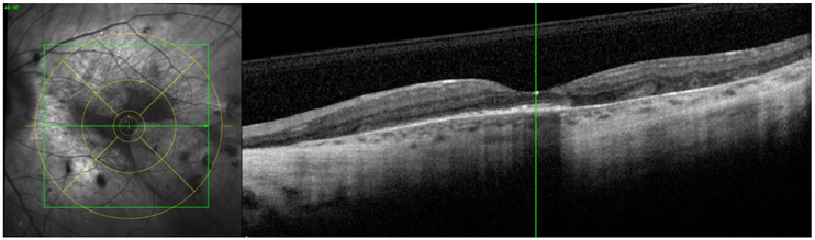
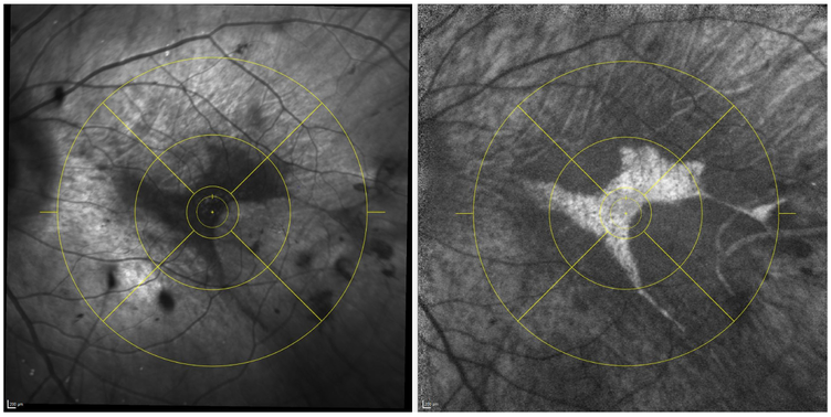
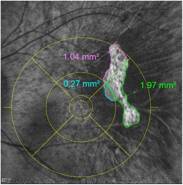
Methods: A cohort of genotypically confirmed choroideremia cases who underwent optical coherence tomography (OCT) and fundus autofluorescence (FAF) imaging was studied. Using HEYEX review software, the foveal centre was marked on FAF images under guidance of corresponding OCT images, followed by application of an ETDRS grid. The boundaries of preserved autofluorescence (AF) were manually segmented in each individual ETDRS subfield. The regional distribution of preserved AF was assessed by comparing its area among the various subfields.
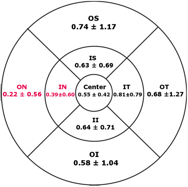
Results: A total of 168 eyes from 84 choroideremia cases were enrolled. There was a statistically significant difference in the amount of preserved AF area between inner subfields as determined by one-way analysis of variance (F (3,668)=9.997, p<0.001) and also between outer subfields (F (3,668)=8.348, p<0.001). A Tukey posthoc test revealed that the preserved AF area in the nasal subfields in both the inner and outer subfields was significantly smaller compared with analogue subfields.
Conclusion: The asymmetric spatial distribution of preserved AF in choroideremia (corresponding to the stellate shaped nature of these regions) suggests that the progression of degeneration has directional preference.
Keywords: choroideremia; ellipsoid zone; fundus autofluorescence; optical coherence tomography; preserved autofluorescence; retinal pigment epithelium.
© Author(s) (or their employer(s)) 2019. No commercial re-use. See rights and permissions. Published by BMJ.
Conflict of interest statement
Competing interests: None declared.
Figures
References
-
- van den Hurk JA, Schwartz M, van Bokhoven H, et al. Molecular basis of choroideremia (CHM): mutations involving the rab escort protein-1 (REP-1) gene. Hum Mutat 1997;9:110–7. - PubMed
-
- Jacobson SG, Cideciyan AV, Sumaroka A, et al. Remodeling of the human retina in choroideremia: rab escort protein 1 (REP-1) mutations. Invest Ophthalmol Vis Sci 2006;47:4113–20. - PubMed
-
- Seabra MC, Brown MS, Goldstein JL. Retinal degeneration in choroideremia: deficiency of rab geranylgeranyl transferase. Science 1993;259:377–81. - PubMed
-
- Cremers FP, van de Pol DJ, van Kerkhoff LP, et al. Cloning of a gene that is rearranged in patients with choroideraemia. Nature 1990;347:674–7. - PubMed