. 2018 Oct 23;115(43):10977-10982.
doi: 10.1073/pnas.1720476115. Epub 2018 Oct 8.

Cryo-ET reveals the macromolecular reorganization of S. pombe mitotic chromosomes in vivo

Affiliations

Cryo-ET reveals the macromolecular reorganization of S. pombe mitotic chromosomes in vivo

Shujun Cai et al. Proc Natl Acad Sci U S A. .

Abstract

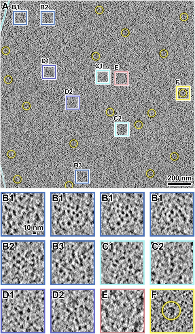
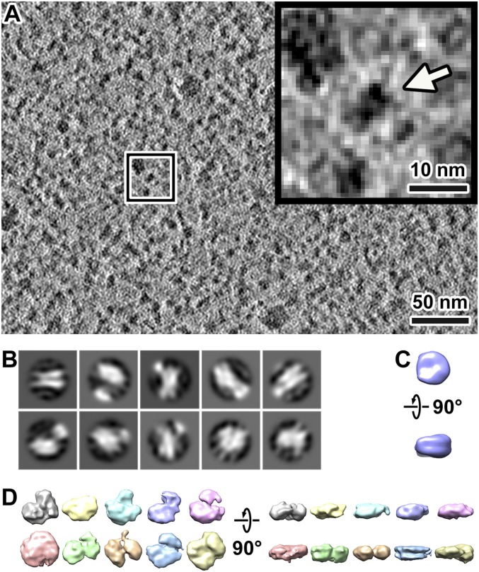
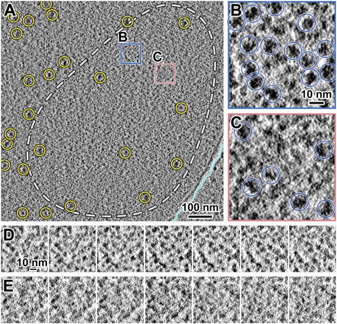
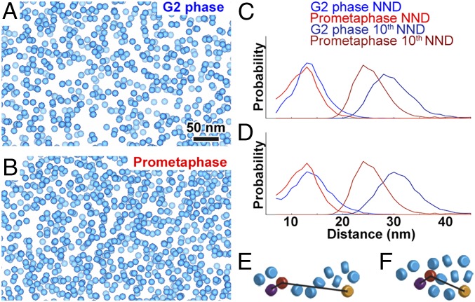
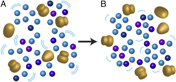
Chromosomes condense during mitosis in most eukaryotes. This transformation involves rearrangements at the nucleosome level and has consequences for transcription. Here, we use cryo-electron tomography (cryo-ET) to determine the 3D arrangement of nuclear macromolecular complexes, including nucleosomes, in frozen-hydrated Schizosaccharomyces pombe cells. Using 3D classification analysis, we did not find evidence that nucleosomes resembling the crystal structure are abundant. This observation and those from other groups support the notion that a subset of fission yeast nucleosomes may be partially unwrapped in vivo. In both interphase and mitotic cells, there is also no evidence of monolithic structures the size of Hi-C domains. The chromatin is mingled with two features: pockets, which are positions free of macromolecular complexes; and "megacomplexes," which are multimegadalton globular complexes like preribosomes. Mitotic chromatin is more crowded than interphase chromatin in subtle ways. Nearest-neighbor distance analyses show that mitotic chromatin is more compacted at the oligonucleosome than the dinucleosome level. Like interphase, mitotic chromosomes contain megacomplexes and pockets. This uneven chromosome condensation helps explain a longstanding enigma of mitosis: a subset of genes is up-regulated.

Keywords: chromatin; condensation; cryo-ET; fission yeast.


Conflict of interest statement

The authors declare no conflict of interest.

Figures

Fig. 1.
Fig. 1.
S. pombe G2-phase chromatin consists of irregularly packed nucleosome-like particles and dispersed megacomplexes. (A) Cryotomographic slice (10 nm) of an interphase nucleus. Yellow circles, a subset of megacomplexes. The cyan lines follow the outer-nuclear membrane. The boxes are enlarged fourfold in BF. (B1B3) Three examples of clusters of nucleosome-like particles. The four B1 panels show left-to-right a series of cryotomographic slices (10 nm) in a step size of 4 nm. (C1 and C2) Two examples of chain-like arrangements of nucleosome-like particles. It is not possible to determine if any of these densities are nucleosomes on the basis of linker DNAs, which generally cannot be seen. (D1 and D2) Two examples of loosely packed nucleosome-like particles. (E) An example position that has few nucleosome-like particles in the center. (F) An example megacomplex (circled). Note that SI Appendix, Fig. S5G shows the 2D-classified nucleosome-like particles reproduce the packing phenomena seen in BE. See Movie S1 for more tomographic slices.
Fig. 2.
Fig. 2.
Condensed S. pombe mitotic chromosomes contain few megacomplexes. (A) Cryotomographic slice (11 nm) of a prometaphase cell. Cyan line, nuclear envelope; yellow circles, megacomplexes. The dashed white line roughly outlines a condensed chromosome. Rectangular boxes are enlarged sixfold in B and C. (B) An example position with many nucleosome-like particles. (C) An example position with a megacomplex and fewer nucleosome-like particles. Yellow circle, a megacomplex. (D and E) Consecutive cryotomographic slices (10 nm) in a 2-nm step size of positions shown in B and C, respectively. See Movie S2 for more tomographic slices.
Fig. 3.
Fig. 3.
Subtomogram averages of nucleosome-like particles do not resemble the canonical nucleosomes. (A) Cryotomographic slice (10 nm) of a G2-phase cell nucleoplasm. (Inset) A nucleosome-like particle, indicated by an arrow, enlarged fivefold from the position boxed in white. (B) The 2D classification of nucleosome template-matching hits from four Volta cryotomograms of G2-phase chromatin. (C) A simulated density map of the canonical nucleosome [PDB 1ID3 (65)], low-pass–filtered to 40-Å resolution. (D) Isosurface renderings of representative 3D class averages of template-matching hits in four Volta cryotomograms of G2-phase chromatin. The particles contributing to each class average are likely to be heterogeneous.
Fig. 4.
Fig. 4.
The macromolecular complexes are unevenly packed within S. pombe mitotic condensed chromosomes. (A) Cryotomographic slice (11 nm) of a nucleus in a prometaphase cell, imaged with Volta phase contrast. White dashed line, approximate chromosome boundary. Yellow circles, megacomplexes. The positions in the blue and salmon boxes are enlarged sixfold in B and C, respectively. (B) A position within the mitotic chromosome that contains many closely packed nucleosome-like particles. (C) A position within the mitotic chromosome that contains fewer, loosely packed nucleosome-like particles. Blue circles, 2D-classified nucleosome-like particles. Notice that the centers of mass of some particles are “above” or “below” the tomographic slice shown. These particles therefore appear either smaller or less dense than the other nucleosome-like particles. (D and E) Consecutive cryotomographic slices (10 nm) in a 2-nm step size of the positions shown in B and C, respectively. See Movie S3 for more tomographic slices.
Fig. 5.
Fig. 5.
Comparison of macromolecular complex packing in G2-phase and prometaphase S. pombe cells. (A and B) Template-matched, 2D-classified filtered hits of nucleosome-like particles rendered as blue spheres in Volta cryotomograms of a (A) G2-phase and (B) prometaphase cell. (C) Nearest-neighbor distance (NND) and 10th NND analysis of template-matching hits. x axis, NND or 10th NND, 1-nm bins; y axis, normalized probability. (D) NND and 10th NND of compression-compensated template-matching hits. (E and F) Cartoons showing how (E) two small or (F) one large cluster of nucleosomes (rounded cylinders) could have similar NND, but different 10th NND. In each panel, the purple and orange nucleosomes are the respective nearest and 10th-nearest neighbors of the red nucleosome.
Fig. 6.
Fig. 6.
A molecular model of interphase and mitotic chromatin. (A) Interphase chromatin is loosely packed, with many megacomplexes and pockets. Blue, purple, and dark blue, nucleosome-like particles, with heterogeneity denoted by the variability in colors; large gold bodies, megacomplexes. Megacomplexes (preribosomes, spliceosomes, transcription-preinitiation complexes, etc.) are interspersed between the nucleosome-like particles and macromolecular-complex–free pockets. (B) During mitotic chromosome condensation, most nucleosome-like particles form large clusters, thereby excluding or inhibiting the assembly of megacomplexes. Furthermore, chromatin dynamics (magnitude proportional to the size and number of the curved lines) also decrease. In both interphase and mitotic chromatin, ordered motifs are largely absent.

References

    1. Dixon JR, Gorkin DU, Ren B. Chromatin domains: The unit of chromosome organization. Mol Cell. 2016;62:668–680. - PMC - PubMed
    1. Luger K, Mäder AW, Richmond RK, Sargent DF, Richmond TJ. Crystal structure of the nucleosome core particle at 2.8 A resolution. Nature. 1997;389:251–260. - PubMed
    1. Ricci MA, Manzo C, García-Parajo MF, Lakadamyali M, Cosma MP. Chromatin fibers are formed by heterogeneous groups of nucleosomes in vivo. Cell. 2015;160:1145–1158. - PubMed
    1. Dixon JR, et al. Topological domains in mammalian genomes identified by analysis of chromatin interactions. Nature. 2012;485:376–380. - PMC - PubMed
    1. Mizuguchi T, et al. Cohesin-dependent globules and heterochromatin shape 3D genome architecture in S. pombe. Nature. 2014;516:432–435. - PMC - PubMed

Publication types

MeSH terms