Review
. 2019 Mar;68(3):547-561.
doi: 10.1136/gutjnl-2018-316906. Epub 2018 Oct 8.

Tight junction proteins in gastrointestinal and liver disease

Affiliations
Review

Tight junction proteins in gastrointestinal and liver disease

Mirjam B Zeisel et al. Gut. 2019 Mar.

Abstract

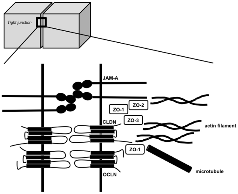
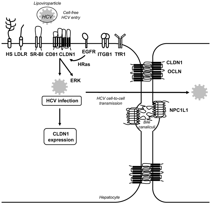
Over the past two decades a growing body of evidence has demonstrated an important role of tight junction (TJ) proteins in the physiology and disease biology of GI and liver disease. On one side, TJ proteins exert their functional role as integral proteins of TJs in forming barriers in the gut and the liver. Furthermore, TJ proteins can also be expressed outside TJs where they play important functional roles in signalling, trafficking and regulation of gene expression. A hallmark of TJ proteins in disease biology is their functional role in epithelial-to-mesenchymal transition. A causative role of TJ proteins has been established in the pathogenesis of colorectal cancer and gastric cancer. Among the best characterised roles of TJ proteins in liver disease biology is their function as cell entry receptors for HCV-one of the most common causes of hepatocellular carcinoma. At the same time TJ proteins are emerging as targets for novel therapeutic approaches for GI and liver disease. Here we review our current knowledge of the role of TJ proteins in the pathogenesis of GI and liver disease biology and discuss their potential as therapeutic targets.

Keywords: colorectal cancer; hepatitis C; hepatocellular carcinoma; tight junction.


Conflict of interest statement

Competing interests: TFB is a coinventor of a patent/patent application of CLDN1-specific antibodies for prevention and treatment of HCV infection. TFB and MBZ are coinventors of patent applications for anti-claudin 1 monoclonal antibodies for the prevention and treatment of liver disease and HCC.

Figures

Figure 1.
Figure 1.. Schematic representation of the expression and function of the major tight junction proteins addressed in this review.
This simplified cartoon only displays the localization and interactions of the major tight junction (TJ) protein families that are addressed in this review. TJs are composed of transmembrane proteins, including different claudins (CLDNs), tight junction-associated marvel proteins (TAMPs, e.g. OCLN), junctional adhesion molecules (JAMs e.g. JAM-A) as well as cytosolic proteins (e.g. ZO-1, -2 and -3), which connect transmembrane components to the cytoskeleton (actin filaments, microtubules). For a more detailed description please refer to reference[1].
Figure 2.
Figure 2.. Schematic representation of differential regulation of tight junction proteins and associated signaling in gastrointestinal cancer.
Signaling and molecular mechanisms that are known to promote neoplastic growth and cancer malignancy include the receptor tyrosine kinase signaling, inflammatory signaling cascades and non-coding RNAs that perturb tight junction (TJs) expression and function. TJ perturbation alters downstream signaling that target important cellular events in epithelial homeostasis, invasion, chronic inflammation and cancer (Zeb-1/E-cadherin, Wnt signaling, MMP9/Notch signaling and Src/PI3K/Akt signaling). Furthermore, disruption of TJs can result in increased permeability to promote translocation of bacteria and luminal antigens, which then activate IL-6/Stat3 signaling to induce carcinogenic processes.
Figure 3.
Figure 3.. Functional roles of CLDN1 as hepatitis C virus entry factor.
CLDN1 is one of the four main hepatitis C virus (HCV) host factors (i.e. SR-BI, CD81, CLDN1 and OCLN) essential for the early steps of HCV infection. Several other host factors (e.g. highly sulphated heparan sulfate (HS), low-density lipoprotein receptor (LDLR), epidermal growth factor receptor (EGFR), integrin beta 1 (ITGB1), transferrin receptor 1 (TfR1) and Niemann Pick C1 like 1 (NPC1L1)) contribute to viral binding and entry. EGFR-mediated signaling leads to the formation of a CD81-CLDN1 co-receptor complex that ultimately leads to viral internalization[116, 118]. HCV infection induces CLDN1-depend signaling via the ERK1/2 pathway[120]. HCV infection increases CLDN1 expression[122, 145]. TJ proteins involved in the HCV entry process are depicted in black, non-TJ host entry factors are depicted in white.
Figure 4.
Figure 4.. Functional role of CLDN1 in signal transduction and EMT in liver disease.
In transformed liver cells, CLDN1 over-expression activates the c-Abl-PKC pathway to increase cellular migration and invasion via MMP2 activation[20] as well as the c-Abl-Ras-Raf-ERK pathway to promote EMT via the transcription factors Slug and Zeb1[162].

References

    1. Zihni C, Mills C, Matter K, et al. Tight junctions: from simple barriers to multifunctional molecular gates. Nat Rev Mol Cell Biol 2016;17:564–80. - PubMed
    1. Singh AB, Uppada SB, Dhawan P. Claudin proteins, outside-in signaling, and carcinogenesis. Pflugers Arch 2017;469:69–75. - PMC - PubMed
    1. Brandner JM, Zorn-Kruppa M, Yoshida T, et al. Epidermal tight junctions in health and disease. Tissue Barriers 2015;3:e974451. - PMC - PubMed
    1. Zeisel MB, Turek M, Baumert TF. Tight junctions and viral entry. Future Virology 2010;5:263–71.
    1. Cummins PM. Occludin: one protein, many forms. Mol Cell Biol 2012;32:242–50. - PMC - PubMed

Publication types

MeSH terms