. 2018 Oct 8;23(10):2559.
doi: 10.3390/molecules23102559.

Identification of Anti-Melanogenesis Constituents from Morus alba L. Leaves

Affiliations

Identification of Anti-Melanogenesis Constituents from Morus alba L. Leaves

Hong Xu Li et al. Molecules. .

Abstract

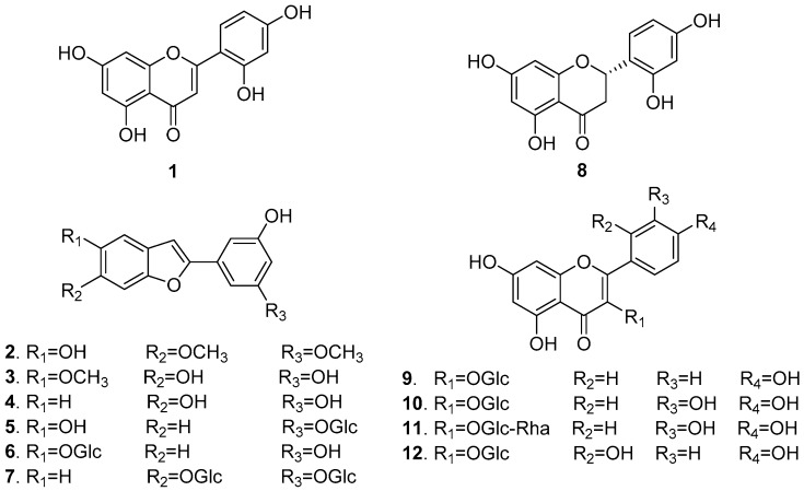
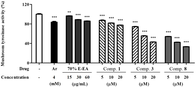
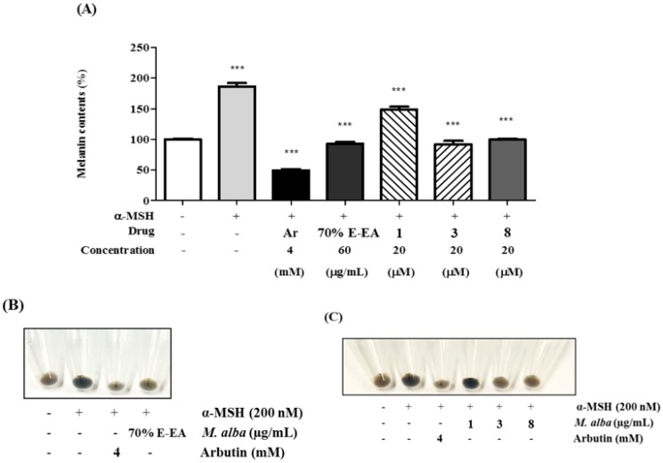
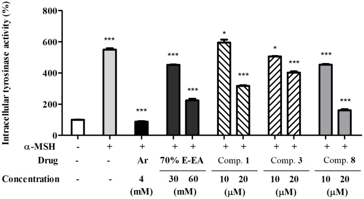
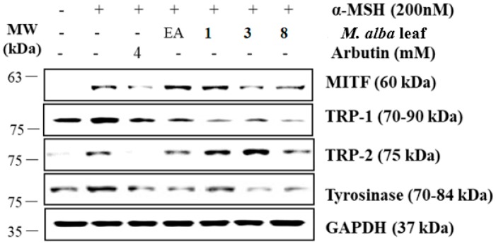
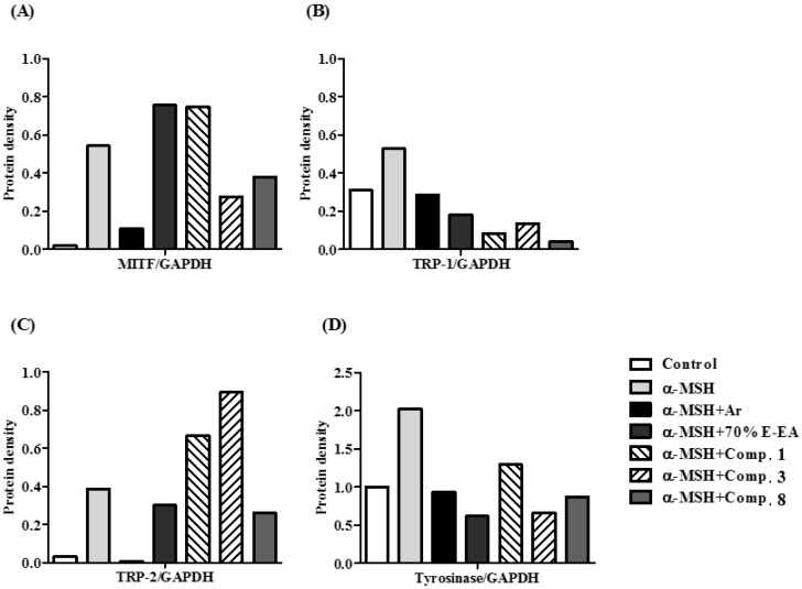
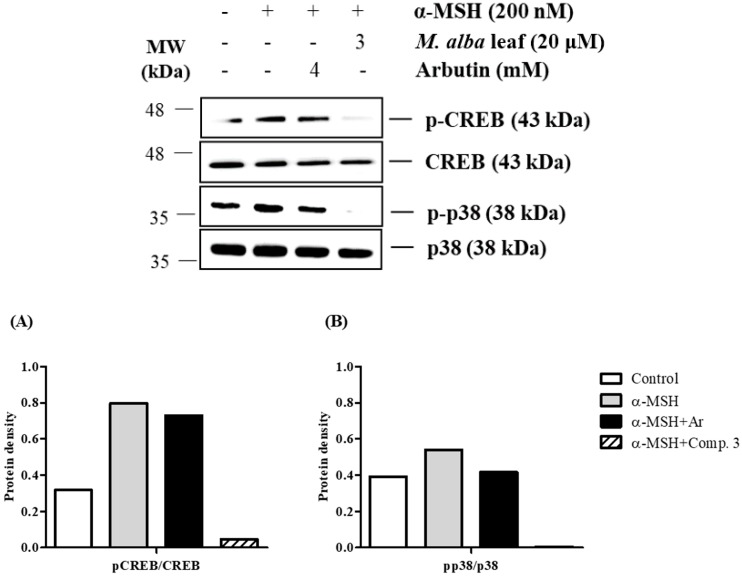
The individual parts of Morus alba L. including root bark, branches, leaves, and fruits are used as a cosmetic ingredient in many Asian countries. This study identified several anti-melanogenesis constituents in a 70% ethanol extract of M. alba leaves. The ethyl acetate fraction of the initial ethanol extract decreased the activity of tyrosinase, a key enzyme in the synthetic pathway of melanin. Twelve compounds were isolated from this fraction and their structures were identified based on spectroscopic spectra. Then, the authors investigated the anti-melanogenesis effects of the isolated compounds in B16-F10 mouse melanoma cells. Compounds 3 and 8 significantly inhibited not only melanin production but also intracellular tyrosinase activity in alpha-melanocyte-stimulating-hormone (α-MSH)-induced B16-F10 cells in a dose-dependent manner. These same compounds also inhibited melanogenesis-related protein expression such as microphthalmia-associated transcription factor (MITF), tyrosinase, and tyrosinase-related protein-1 (TRP-1). Compound 3 modulated the cAMP-responsive element-binding protein (CREB) and p38 signaling pathways in α-MSH-activated B16-F10 melanoma cells, which resulted in the anti-melanogenesis effects. These results suggest that compound 3, isolated from M. alba leaves, could be used to inhibit melanin production via the regulation of melanogenesis-related protein expression.

Keywords: MITF; Morus alba leaf; TRP-1; TRP-2; anti-melanogenesis; tyrosinase.


Conflict of interest statement

The authors declare no conflict of interest.

Figures

Figure 1
Figure 1
Structures of compounds 112 from M. alba leaves.
Figure 2
Figure 2
Inhibitory effects of M. alba leaves on mushroom tyrosinase activity; a mushroom tyrosinase activity was assayed by measuring 3,4-dihydroxyphenylalanine (DOPA) oxidase activity. The test samples were diluted in sodium phosphate buffer (67 mM, pH 6.8) and added to the samples in 96-well plates. Then, l-dopa (2 mM) and mushroom tyrosinase (200 U) were added to each well for 10 min. The absorbance was measured at 490 nm by using an ELISA microplate reader. Results are expressed as percentages of ** p < 0.01 and *** p < 0.001 compared with the control group. 70% E-EA indicates ethyl acetate fraction from 70% ethanol extract of M. alba leaves.
Figure 3
Figure 3
Effects on melanogenesis in murine B16-F10 cells; B16-F10 cells were pretreated with test samples for 2 h and then incubated with alpha-melanocyte-stimulating-hormone (α-MSH) (200 nM) for 72 h. The cell pellets were dissolved in 1N NaOH with 10% DMSO. The cell lysates were transferred to 96-well plates and the absorbances at 405 nm were detected using an ELISA microplate reader. (A) 70% E-EA, 1, 3, and 8 decreased the α-MSH-induced production of melanin. *** p < 0.001 compared with the α-MSH-treated group; (B,C) indicate the colors of the pellets from α-MSH-induced B16-F10 melanoma cells.
Figure 4
Figure 4
Effects on intracellular tyrosinase activity in murine B16-F10 cells; B16-F10 cells were pretreated with test samples for 2 h and then incubated with α-MSH (200 nM) for 72 h. The cell lysates (50 μg) were dissolved in 100 mM phosphate buffer (pH 6.8) and treated with L-dopa (2 mg/mL) in a 96-well plate at 37 °C for 2 h. The production amount of dopachrome was measured by using an ELISA microplate reader at 490 nm. * p < 0.05 and *** p < 0.001 compared with the α-MSH-treated group.
Figure 5
Figure 5
Effects on melanogenesis-related protein expression in α-MSH-induced murine B16-F10 cells; B16-F10 cells were pretreated with test samples for 2 h and α-MSH was added to the cells for 24 h. Glyceraldehyde 3-phosphate dehydrogenase (GAPDH) was used as the control protein. The protein levels of microphthalmia-associated transcription factor (MITF), tyrosinase, tyrosinase-related protein-1 (TRP-1), and tyrosinase-related protein-2 (TRP-2) were detected by Western blot analysis. Compounds 3 and 8 significantly reduced the expression of MITF, tyrosinase, TRP-1, and TRP-2 in α-MSH-induced B16-F10 cells. Arbutin, a positive control drug, also blocked the melanogenesis. (A) MITF, (B) TRP-1, (C) TRP-2, and (D) tyrosinase were analyzed and quantified using Azure Spot software. MITF, TRP-1, TRP-2 and tyrosinase protein levels were normalized by GAPDH.
Figure 5
Figure 5
Effects on melanogenesis-related protein expression in α-MSH-induced murine B16-F10 cells; B16-F10 cells were pretreated with test samples for 2 h and α-MSH was added to the cells for 24 h. Glyceraldehyde 3-phosphate dehydrogenase (GAPDH) was used as the control protein. The protein levels of microphthalmia-associated transcription factor (MITF), tyrosinase, tyrosinase-related protein-1 (TRP-1), and tyrosinase-related protein-2 (TRP-2) were detected by Western blot analysis. Compounds 3 and 8 significantly reduced the expression of MITF, tyrosinase, TRP-1, and TRP-2 in α-MSH-induced B16-F10 cells. Arbutin, a positive control drug, also blocked the melanogenesis. (A) MITF, (B) TRP-1, (C) TRP-2, and (D) tyrosinase were analyzed and quantified using Azure Spot software. MITF, TRP-1, TRP-2 and tyrosinase protein levels were normalized by GAPDH.
Figure 6
Figure 6
Effects of M. alba leaves on the phosphorylation of the cAMP-responsive element-binding protein (CREB) and p38 in α-MSH-induced B16-F10 cell; B16-F10 cells were pretreated with test samples for 2 h and α-MSH was added to the cells for 24 h. Total protein was used as the control protein. The protein levels of pCREB, CREB, pp38, p38 were detected by Western blot analysis. Compound 3 significantly reduced the expression of pCREB and pp38 in α-MSH-induced B16-F10 cells. Arbutin, a positive control drug, also blocked the melanogenesis. The protein level of (A) pCREB and (B) pp38 were quantitated with Azure Spot software. pCREB and pp38 protein levels were normalized by the total protein of CREB or p38.

References

    1. Costin G.E., Hearing V.J. Human skin pigmentation: Melanocytes modulate skin color in response to stress. FASEB J. 2007;21:976–994. doi: 10.1096/fj.06-6649rev. - DOI - PubMed
    1. D’Mello S.A., Finlay G.J., Baguley B.C., Askarian-Amiri M.E. Signaling pathways in melanogenesis. Int. J. Mol. Sci. 2016;17:1144. doi: 10.3390/ijms17071144. - DOI - PMC - PubMed
    1. Lin J.Y., Fisher D.E. Melanocyte biology and skin pigmentation. Nature. 2007;445:843. doi: 10.1038/nature05660. - DOI - PubMed
    1. Park H., Kosmadaki M., Yaar M., Gilchrest B. Cellular mechanisms regulating human melanogenesis. Cell. Mol. Life Sci. 2009;66:1493–1506. doi: 10.1007/s00018-009-8703-8. - DOI - PMC - PubMed
    1. Slominski A., Tobin D.J., Shibahara S., Wortsman J. Melanin pigmentation in mammalian skin and its hormonal regulation. Physiol. Rev. 2004;84:1155–1228. doi: 10.1152/physrev.00044.2003. - DOI - PubMed

MeSH terms