. 2018 Oct 8;8(1):14934.
doi: 10.1038/s41598-018-33355-5.

Temperature affects the biology of Schmidtea mediterranea

Affiliations

Temperature affects the biology of Schmidtea mediterranea

Nassim Hammoudi et al. Sci Rep. .

Abstract

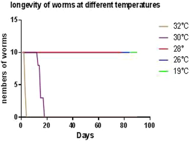
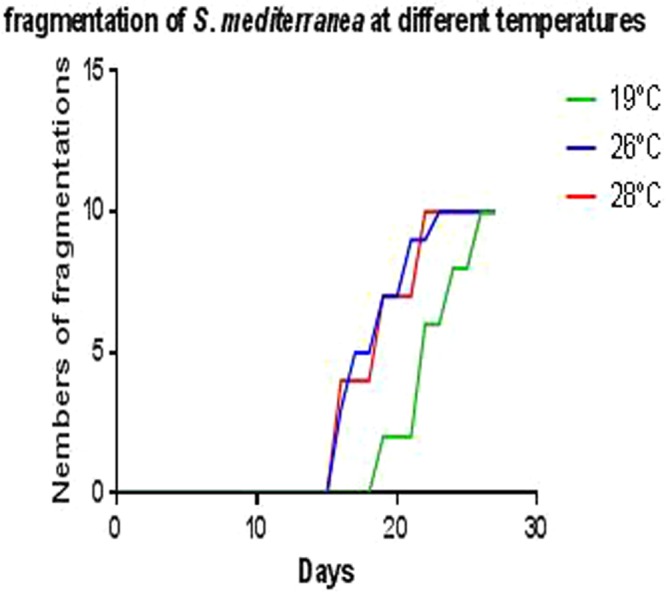
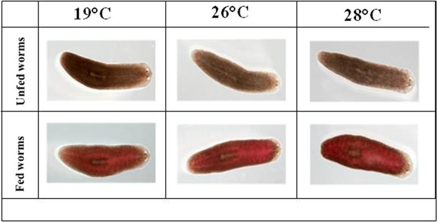
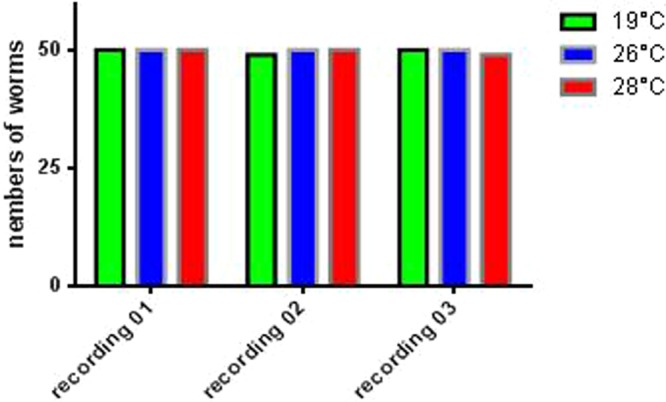
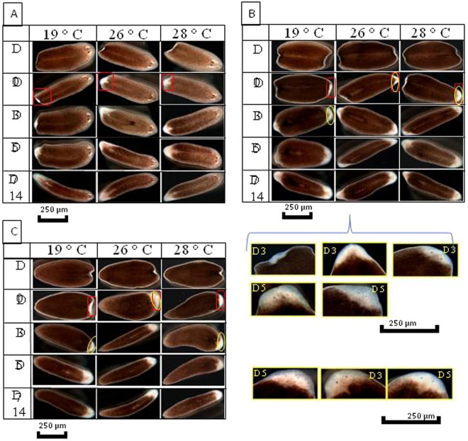
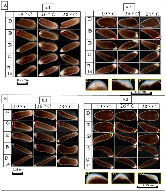
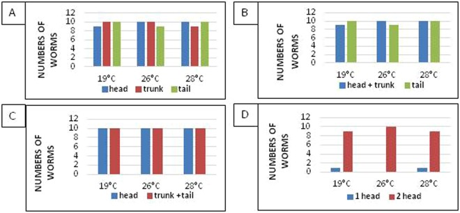
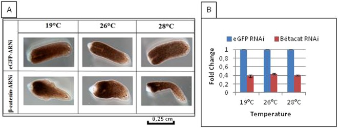
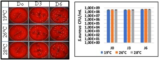
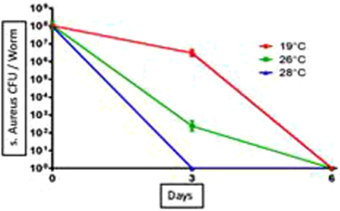
Studies of tissue regeneration and host-pathogen interactions using the model planarian Schmidtea mediterranea have been performed at an experimental temperature of 19 °C. S. mediterranea planarians exposed to 19 °C-32 °C were observed for survival, mobility, feeding and regeneration for three months and elimination of the Staphylococcus aureus pathogen over six days. S. mediterranea planarians died at 30 °C-32 °C after 18 days of observation but tolerated temperatures of 19 °C up to 28 °C with non-significant differences in mobility and feeding behavior. Genetic malleability tested by RNAi feeding was still efficient at 26 °C and 28 °C. Concerning the immune capacity of planarians, we reported an exacerbation of the immune response in worms infected by S. aureus at 26 °C and 28 °C. These observations suggest a temperature modulation of planarian stem cells and illustrate the importance of modulating experimental temperature when using planarians as model organisms to study regeneration and immune response.


Conflict of interest statement

The authors declare no competing interests.

Figures

Figure 1
Figure 1
Longevity of the planarians incubated at different temperatures. The planarians exposed to a temperature increase of 1 °C every 25 min, a daily monitoring of their survival allowed us to observe the death of the worms incubated at 32 °C and 30 °C after 4 and 18 days respectively, the planarians incubated at 26 °C and 28 °C remain alive after three months of incubation.
Figure 2
Figure 2
Spontaneous fragmentation of planarians incubated at different temperature. Follow-up of the number of spontaneous fragmentations observed in planarians incubated at different temperatures reveals a beginning of fragmentation from the 16th day for the worms incubated at 26 °C and 28 °C, this is observed on the 19th day at 19 °C. A doubling of the number of worms is observed after 23 days at 26 °C and 28 °C and it was not until the 26th day that it was reported at 19 °C.
Figure 3
Figure 3
Mobility of the planarians incubated at different temperature. The biostatistical analysis with the BiostaTGV software of the results obtained from the videos of mobility of the worms allowed us to calculate the average speeds of movements in each realized film. This revealed to us 0.65 × 10−5 m/s, 0.7 × 10−5 m/s, and 0.68 × 10−5 ± 0.02 m/s (p = 0.06) at 19 °C, 26 °C, 28 °C respectively.
Figure 4
Figure 4
Presentation of phenotypes of fed planarians and incubated at different temperatures, namely 19, 26, 28 °C.
Figure 5
Figure 5
Feeding behavior of the planarians incubated at different temperature. Three planarian groups (n = 50) adapted to 19, 26, 28 °C, are fed three consecutive times at an interval of 2 days to estimate their aptitude to eat, at 19 °C there were 50, 49, 50 ± 0.5773 fed, at 26 °C 50 worms fed in all experiments, finally, at 28 °C, 50, 50, 49 ± 0.5773 worms fed.
Figure 6
Figure 6
Intensity of coloration after the feeding of the planarians incubated at different temperatures. The feeding of the worms is followed by a calculation of the color intensity of the worms using the ImageJ software, this analysis shows us a non-significant difference in the color intensity of the worms incubated in different temperatures. Indeed, 1.97, 1.98, 1.96 (UAC) were obtained at 19, 26, 28 °C respectively.
Figure 7
Figure 7
Regeneration of planarians after amputation of the head and tail. (A) Regeneration of the head: appearance of the blastema at Day 3 on all the fragments, the blastema formation is complete after Day 7, and the regeneration ends at Day 14. (B) Regeneration of the trunk: we see the appearance of the blastema on Day 3 with the formation of the eyes on the worms incubated at 26 °C, concerning the worms incubated at 19 and 28 °C the eyes are formed on Day 5. (C) Regeneration of the trunk: at Day 3 post-amputation, we see the formation of blastema on all the fragments at different temperatures, this is followed by the appearance of the eyes in worms incubated at 26 °C; at 19 °C and 28 °C, the eyes only appear at Day 5. The complete formation of the blastema is made on Day 7 and the completion of the regeneration process is observed on Day 14.
Figure 8
Figure 8
(A) Regeneration of planarians after amputation of the head. (a.1) Regeneration of the head: appearance of the blastema on all the fragments on Day 3, then completely formed on Day 7, and the regeneration ends on Day 14. (a.2) Regeneration of the trunk-tail: formation of the regeneration blastema on Day 3 and an appearance of the eyes in worms incubated at 26 °C is reported, however, at 19 °C and 28 °C the eyes do not appear until Day 5. (B) Regeneration of planarians after amputation of the tail. (b.2) Regeneration of the head-trunk: after the section of the tail, the formation of blastema begins on Day 3 and ends on Day 7 for all the fragments, and complete worms are formed on Day 14. (b.2) Regeneration of the tail: characterized by the appearance of the eyes on blastema of worms incubated at 26 and 28 °C, this observation is reported on Day 5 for worms incubated at 19 °C.
Figure 9
Figure 9
Quantification of the different phenotypes observed after fragmentation. (A) Number of worms regenerated after three cuts (head, trunk, tail) with a P = 1 according to temperature. (B) Number of worms regenerated after two cuts (head + trunk, tail) P = 0.64 according to temperature. (C) Number of worms regenerated after two cuts (head, trunk + tail) P = 0.46. (D) Number of worms with one or two heads after invalidation of β-catenin gene as a function of temperature P = 1.
Figure 10
Figure 10
Regeneration of the planarian head-trunk part invalidated for the eGFP or β-catenin genes. (A) After the genes were invalidated, the worms were cut. At day 5 of the regeneration, there was appearance of the head instead of the tail in worms invalidated for the β-catenin gene, a normal regeneration in worms invalidated for the gene eGFP (jellyfish gene non-existent in the Planarian genome). (B) The fold change of the β-catenin and eGFP genes invalidated in 10 planarians incubated in the three different temperatures (19 °C, 26 °C, 28 °C) reveals a non-significant difference with P = 0.99.
Figure 11
Figure 11
Test viability of S. aureus incubated in water at different temperatures. After incubation of the bacteria in water at 19 °C, 26 °C and 28 °C the bacteria are enumerated every 3 days by counting the CFUs. Results showed a non-significant difference as a function of the incubation temperature with P = 0.99.
Figure 12
Figure 12
Kinetics of elimination of S. aureus by planarians. After infection of the planarians with S. aureus, the bacteria were enumerated every 3 days by counting the CFUs. The elimination kinetics revealed a clearance at Day 6 post-infection at 19 °C and 26 °C, however this clearance was observed on Day 3 in worms incubated at 28 °C. Elimination of bacteria by planarians was faster at 28 °C and 26 °C compared to control 19 °C. The results are expressed as mean ± standard deviation (10 worms per point, n = 3, *p < 0.05). The results are analyzed with GraphPad Prism© software using the non-parametric Mann-Whitney U test.
Figure 13
Figure 13
Reproducibility test of the results of the elimination of S. aureus by the planarians. (AC) Represent five replicates of S. aureus eliminated by the planarians in each incubation condition 19 °C, 26 °C and 28 °C, showing non-significant differences with P = 0.89; 0.58; 0.99, respectively.
Figure 14
Figure 14
Fragmentation of planarians. (A) Fragmentation of the planarians in three parts head, trunk, tail. (B) Amputation of the head. (C) Amputation of the tail (ref.).

References

    1. Collins JJ. Platyhelminthes. Curr. Biol. 2017;27:R252–R256. doi: 10.1016/j.cub.2017.02.016. - DOI - PubMed
    1. Roberts-Galbraith RH, Newmark PA. On the organ trail: Insights into organ regeneration in the planarian. Curr. Opin. Genet. Dev. 2015;32:37–46. doi: 10.1016/j.gde.2015.01.009. - DOI - PubMed
    1. Benedict, J. E. et al. P. S. Pallas medicinae doctoris Miscellanea zoologica: quibus novae imprimis atque obscurae animalium species describuntur et observationibus iconibusque illustrantur, 10.5962/bhl.title.69851 (Apud Petrum van Cleef, 1766).
    1. Morgan TH. Experimental studies of the regeneration of Planaria maculata. Arch. für Entwickelungsmechanik der Org. 1898;7:364–397. doi: 10.1007/BF02161491. - DOI
    1. Alvarado AS, Tsonis PA. Bridging the regeneration gap: genetic insights from diverse animal models. Nat. Rev. Genet. 2006;7:873–884. doi: 10.1038/nrg1923. - DOI - PubMed