. 2018 Oct;24(10):1513-1518.
doi: 10.1038/s41591-018-0184-6. Epub 2018 Oct 8.

In utero CRISPR-mediated therapeutic editing of metabolic genes

Affiliations

In utero CRISPR-mediated therapeutic editing of metabolic genes

Avery C Rossidis et al. Nat Med. 2018 Oct.

Abstract

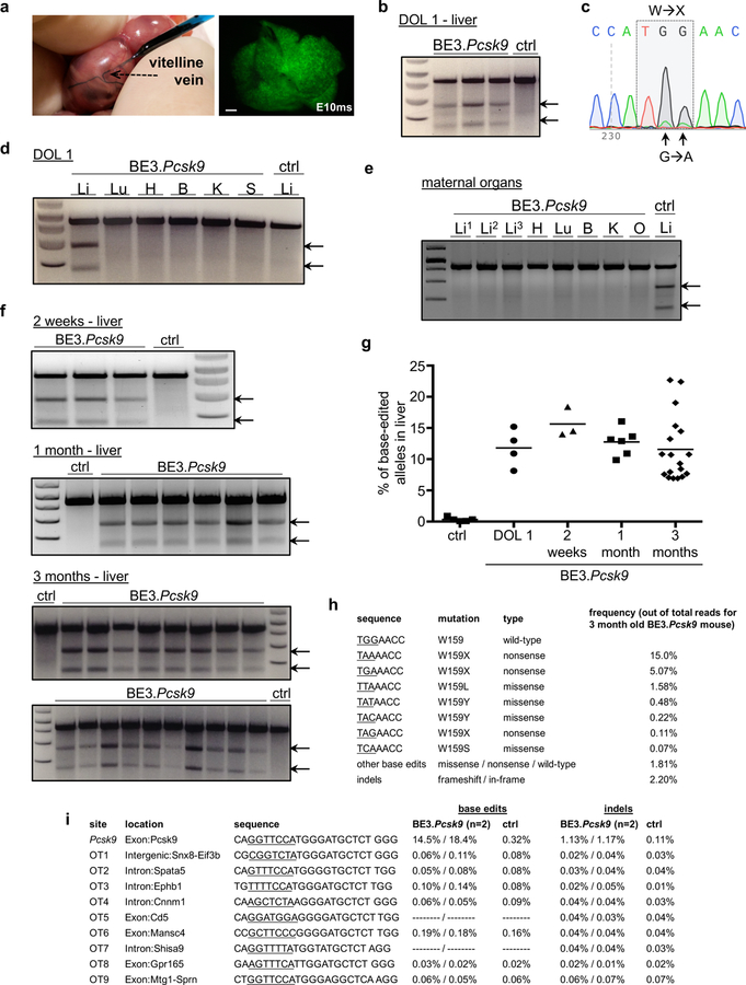
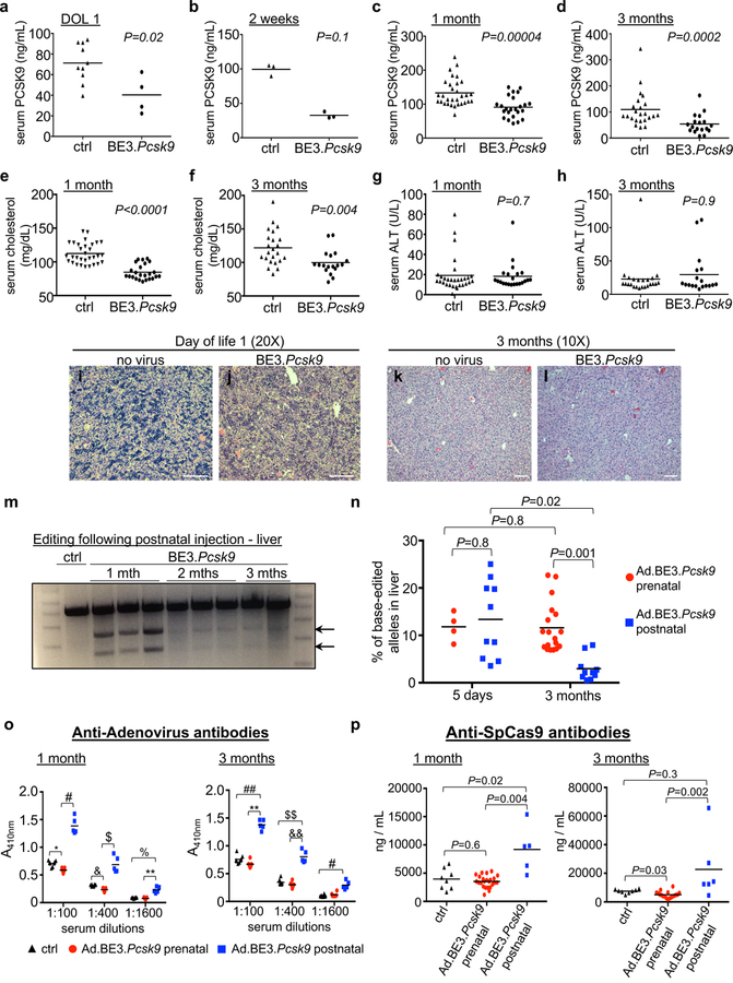
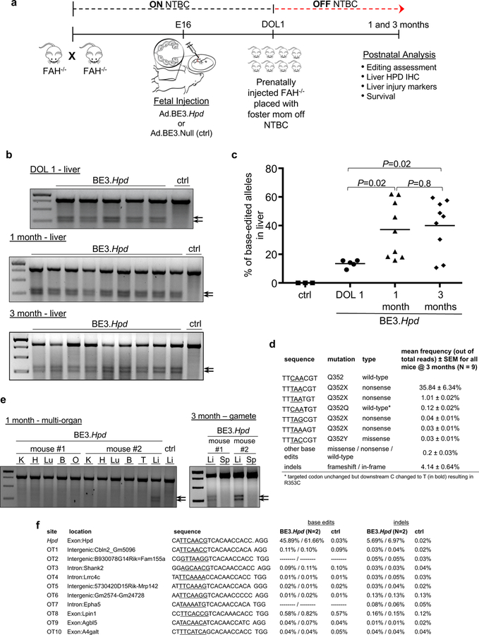
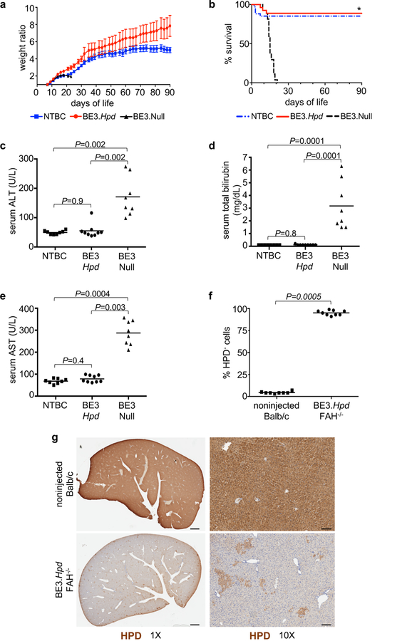
In utero gene editing has the potential to prenatally treat genetic diseases that result in significant morbidity and mortality before or shortly after birth. We assessed the viral vector-mediated delivery of CRISPR-Cas9 or base editor 3 in utero, seeking therapeutic modification of Pcsk9 or Hpd in wild-type mice or the murine model of hereditary tyrosinemia type 1, respectively. We observed long-term postnatal persistence of edited cells in both models, with reduction of plasma PCSK9 and cholesterol levels following in utero Pcsk9 targeting and rescue of the lethal phenotype of hereditary tyrosinemia type 1 following in utero Hpd targeting. The results of this proof-of-concept work demonstrate the possibility of efficiently performing gene editing before birth, pointing to a potential new therapeutic approach for selected congenital genetic disorders.


Conflict of interest statement

COMPETING FINANCIAL INTERESTS

The authors have no conflicts of interest to declare.

Figures

Figure 1
Figure 1
In utero base editing of the Pcsk9 gene. (a) Vitelline vein injection of E16 fetus; DOL1 liver, Ad.GFP injection (fluorescence stereomicroscopy, GFP filter). Scale bar = 1mm. (b-i) E16 Balb/c fetuses were injected with Ad.BE3.Pcsk9 or Ad.BE3.Null (ctrl). Genomic DNA from organs of injected fetuses (b-d; f-i) or of dams of injected fetuses (e) were assessed for Pcsk9 on-target (b-h) and off-target (i) editing by Surveyor assays (b, d-f), Sanger sequencing (c), and NGS (g-i). Maternal organ analysis (e) occurred 1 week after fetal injection, 3 separate liver samples per mother, ctrl Li = liver DNA from injected fetus. (b, N=3 BE3.Pcsk9, 1 ctrl; c, representative of 4 mice replicates; d, representative of 3 mice replicates; e, representative of 2 mice replicates; f, 2 weeks: N=3 BE3.Pcsk9, 1 ctrl, 1 month: N=6 BE3.Pcsk9, 1 ctrl, 3 months: N=18 BE3.Pcsk9, 2 ctrl) (g) NGS, liver DNA in control mice (N=5) and at DOL1 (N=4), 2 weeks (N=3), 1 month (N=6), and 3 months of age (N=18) following in utero Ad.BE3.Pcsk9 injection; measure of centre = mean. (h) Frequencies of base-edited and indel-bearing alleles, liver DNA, 3-month-old prenatal recipient of Ad.BE3.Pcsk9. Underlined bases indicate the target codon. (i) Base editing and indel rates for the on-target and top 9 predicted off-target sites, NGS, liver DNA at 2 weeks of age from 2 prenatal Ad.BE3.Pcsk9 recipients (results separated by dashes) and a control mouse. Underlined bases indicate the base editing window based on distance from the PAM. In cases in which no base editing rates are shown, there were no C bases within the window. DOL, day of life; BE3, base editor 3; PCSK9, proprotein convertase subtilisin/kexin type 9; ctrl, control; exp; Li, liver; Lu, lung; H, heart; B, brain; K, kidney; S, spleen; arrows, Surveyor cleavage products.
Figure 2
Figure 2
Functional effects of in utero Pcsk9 base editing and comparison to postnatal editing. (a-l) E16 Balb/c fetuses were injected with Ad.BE3.Pcsk9 or Ad.BE3.Null (ctrl). (a-d) Plasma PCSK9 protein levels at DOL1 (ctrl, N=10; BE3.Pcsk9, N=4), 2 weeks (ctrl, N=3; BE3.Pcsk9, N=3), 1 month (ctrl, N=31; BE3.Pcsk9, N=24), and 3 months (ctrl, N=23; BE3.Pcsk9, N=18). (e,f) Plasma cholesterol levels at 1 (ctrl, N=31; BE3.Pcsk9, N=24) and 3 months (ctrl, N=23; BE3.Pcsk9, N=18). (g,h) Plasma ALT levels at 1 (ctrl, N=30; BE3.Pcsk9, N=22) and 3 months (ctrl, N=23; BE3.Pcsk9, N=18). (i-l) Liver histology (hematoxylin and eosin staining) from non-injected and Ad.BE3.Pcsk9 injected fetuses. Representative of 3 mice replicates per group per time point. Scale bar = 100μm (m) Ad.BE3.Pcsk9 was injected into 5-week-old B6 mice. Surveyor assays, liver genomic DNA at 1 month (N=3), 2 months (N=3), and 3 months (N=2) post-injection. (n) Ad.BE3.Pcsk9 was injected into E16 Balb/c fetuses and 5-week-old Balb/c mice. Percentage base-edited Pcsk9 on-target alleles, NGS, liver genomic DNA at 5 days and 3 months post-injection (prenatal 5 days-N=4, 3 months-N=18; postnatal 5 days-N=10, 3 months-N=10). (o,p) Serum from mice injected at E16 or 5 weeks of age with Ad.BE3.Psck9 was assessed at 1 and 3 months post injection for antibodies to adenovirus (o) and SpCas9 (p) (anti-Ad: prenatal 1 month-N=11, 3 months-N=6; postnatal 1 month-N=5, 3 months-N=5; anti-SpCas9: prenatal 1 month-N=24, 3 months-N=18; postnatal 1 month-N=5, 3 months-N=6); Control non-injected, age-matched Balb/c mice (N=8). DOL, day of life; BE3, base editor 3; PCSK9, proprotein convertase subtilisin/kexin type 9; ALT, alanine aminotransferase; arrows, Surveyor cleavage products; * P=0.01; # P=0.001; $ P=0.00008; & P=0.006; % P=0.03; ** P=0.0007; ## P=0.04; $ $ P=0.02; && P=0.002. Statistical analysis performed with two-tailed Mann-Whitney U test (a-d,g,h), two-tailed Student’s t test (e, t=7.8, df=53; f, t=3.2; df=39) and Kruskal-Wallis test (n-p). Measure of centre = mean (a-h, n-p).
Figure 3
Figure 3
In utero base editing of Hpd in the Fah–/– mouse model. (a) Experimental scheme. (b) Surveyor assays to assess Hpd base editing, liver genomic DNA at DOL1, 1 month, and 3 months of age (Ad.BE3.Hpd DOL1, N=5; 1 month, N=9; 3 months, N=9) or Ad.BE3.Null (ctrl, N=1). (c) The percentage of base-edited Hpd on-target alleles was assessed by NGS of liver genomic DNA in fetal recipients of Ad.BE3.Null (ctrl, N=3 at DOL1 to 2 weeks of age) and Ad.BE3.Hpd at DOL1 (N=5), 1 month (N=9), and 3 months of age (N=9). Measure of centre = mean. (d) Frequencies of base-edited and indel-bearing alleles were assessed at 1 month of age via NGS of liver genomic DNA of prenatal Ad.BE3.Hpd recipients (N=9). Underlined bases indicate the target codon. (e) Genomic DNA isolated from other organs at 1 month of age (two independent mice as shown) and sperm at 3 months of age (2 independent mice as shown) was assessed by Surveyor assays for Hpd editing following prenatal Ad.BE3.Hpd injection. – ctrl = liver DNA from Ad.BE3.Null-injected fetus. (f) NGS analysis of the Hpd on-target site and the top 10 predicted off-target sites in liver genomic DNA harvested at 1 month of age from 2 Ad.BE3.Hpd recipients and 1 Ad.BE3.Null (ctrl) recipient. BE3, base editor; HPD, hydroxyphenylpyruvate dioxygenase; NTBC, 2-(2-nitro-4-trifluoro-methylbenzyol)-1,3 cyclohexanedione, FAH, fumarylacetoacetate hydrolase; IHC, immunohistochemistry; DOL, day of life; K, kidney; H, heart; Lu, lung; B, brain; O, ovary; T, testis; L, liver; arrows, Surveyor cleavage products. Statistical analysis performed with Kruskal-Wallis test.
Figure 4
Figure 4
In utero Hpd base editing improves liver function and rescues the lethal phenotype of Fah–/– mice. (a, b) Fah–/– mice injected prenatally with Ad.BE3.Hpd (N=26) or Ad.BE3.Null (N=27) and taken off NTBC at DOL1 and non-injected Fah–/– mice maintained on NTBC (N=33) were serially weighed and followed for survival. Weight ratio represented as mean ± standard error of mean. Survival statistical analysis performed with log-rank test; * P=8×10⁻¹¹ for BE3.Hpd vs. BE3.Null. (c-e) Plasma ALT, total bilirubin, and AST levels were assessed at 1 month of age (or just prior to death in Ad.BE3.Null-injected mice) in Fah–/– mice injected prenatally with Ad.BE3.Hpd (N=9) or Ad.BE3.Null (N=8) and taken off NTBC at DOL1 and non-injected Fah–/– mice maintained on NTBC (N=8). (f, g) Livers of Fah–/– mice prenatally injected with Ad.BE3.Hpd and taken off NTBC at DOL1 (N=9) and non-injected Balb/c mice (N=8) were assessed for HPD staining at 1 month of age. ~100,000 to 300,000 hepatocytes were assessed per sample and the percentages of HPD-negative cells determined. Scale bar = 1mm (g, left panels) and 100μm (g, right panels). Measure of centre = mean (a, c-f). ΒΕ3, base editor; HPD, hydroxyphenylpyruvate dioxygenase; NTBC, 2-(2-nitro-4-trifluoro-methylbenzyol)-1,3 cyclohexanedione; FAH, fumarylacetoacetate hydrolase; ALT, alanine aminotransferase; AST, aspartate aminotransferase. Statistical analysis performed with Kruskal-Wallis test (c-e) and two-tailed Mann-Whitney U test (f).

Comment in

References

    1. Davey MG et al. Induction of immune tolerance to foreign protein via adeno-associated viral vector gene transfer in mid-gestation fetal sheep. PLoS One 12, e0171132 (2017). - PMC - PubMed
    1. Sabatino DE et al. Persistent expression of hF.IX After tolerance induction by in utero or neonatal administration of AAV-1-F.IX in hemophilia B mice. Mol. Ther 15, 1677–1685 (2007). - PubMed
    1. Mingozzi F et al. CD8(+) T-cell responses to adeno-associated virus capsid in humans. Nat. Med 13, 419–422 (2007). - PubMed
    1. Moss RB et al. Repeated aerosolized AAV-CFTR for treatment of cystic fibrosis: a randomized placebo-controlled phase 2B trial. Hum. Gene Ther 18, 726–732 (2007). - PubMed
    1. Endo M et al. The developmental stage determines the distribution and duration of gene expression after early intra-amniotic gene transfer using lentiviral vectors. Gene Ther 17, 61–71 (2010). - PubMed

Publication types