

Comparison of the Techniques of Secondary Intraocular Lens Implantation after Penetrating Keratoplasty
- PMID: 30298105
- PMCID: PMC6157166
- DOI: 10.1155/2018/3271017
Comparison of the Techniques of Secondary Intraocular Lens Implantation after Penetrating Keratoplasty
Abstract
Aim: To conduct a retrospective analysis of secondary IOL implantation in patients who underwent PK with no simultaneous IOL implantation.
Materials and methods: The retrospective study of the secondary implantation of IOLs was conducted in 46 eyes that underwent a primary operation with PK and cataract/lens extraction with no IOL implantation due to capsule rupture or combining corneal or intraocular complications. The minimum period from PK was 12 months. All secondary IOL implantations were performed from January 2011 to August 2017. Aphakic postkeratoplasty patients were treated using one of the surgical techniques for secondary IOL implantation. In-the-bag IOL implantation was possible if the posterior capsule was complete. If the lens capsule remnants were sufficient to provide secure IOL support, an in-the-sulcus IOL implantation was performed. Scleral fixation was offered in eyes with extensive capsular deficiency or the presence of the vitreous body in anterior chamber. BCVA and expected and achieved refraction were evaluated; we included using two biometry devices, and results were compared.
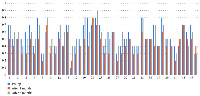
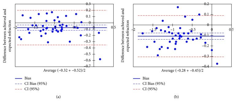
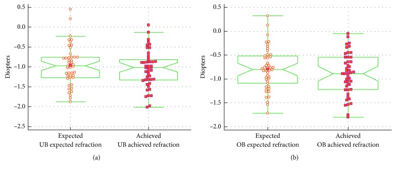
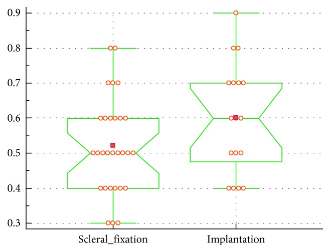
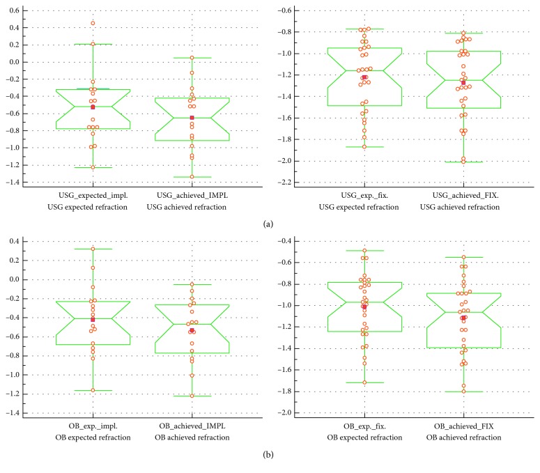
Results: The corrected distance visual acuity (CDVA) before surgery ranged from 0.1 to 0.8 (mean 0.54 ± 0.17). After secondary IOL implantation, CDVA ranged from 0.2 to 0.8 (mean 0.43 ± 0.14) at postoperative 1 month and from 0.3 to 0.9 (mean 0.55 ± 0.15) at postoperative 6 months (p < 0.05). Comparison of the final refraction using two methods of biometry showed no statistically significant difference in the group that underwent scleral fixation of the IOL, similar to the findings for the in-the-bag and in-the-sulcus IOL implantation groups. In the scleral-fixation group, p=0.55 for the USG biometry technique and p=0.22 for the OB technique. p values for the IOL-implantation group were p=0.49 and p=0.44, respectively.
Conclusion: Both implantation methods are safe for the patients. Final refraction is depending on the technique and indication to keratoplasty. Both biometry techniques deliver precise data for IOL choice.
Figures
Similar articles
-
Comparison of Artisan iris-claw intraocular lens implantation and posterior chamber intraocular lens sulcus fixation for aphakic eyes.Int J Ophthalmol. 2014 Apr 18;7(2):283-7. doi: 10.3980/j.issn.2222-3959.2014.02.16. eCollection 2014. Int J Ophthalmol. 2014. PMID: 24790871 Free PMC article.
-
[Comparison of effects of secondary in-the-bag and sulcus intraocular lens implantation in pediatric aphakia after congenital cataract operation].Zhonghua Yan Ke Za Zhi. 2013 Aug;49(8):700-5. Zhonghua Yan Ke Za Zhi. 2013. PMID: 24246808 Chinese.
-
Comparison of secondary implantation of flexible open-loop anterior chamber and scleral-fixated posterior chamber intraocular lenses.J Cataract Refract Surg. 2003 Feb;29(2):301-8. doi: 10.1016/s0886-3350(02)01526-2. J Cataract Refract Surg. 2003. PMID: 12648641
-
[Sutureless scleral intraocular lens fixation: report of nine cases and literature review].J Fr Ophtalmol. 2013 Oct;36(8):658-68. doi: 10.1016/j.jfo.2012.09.009. Epub 2013 Jul 25. J Fr Ophtalmol. 2013. PMID: 23891322 Review. French.
-
Posterior capsulorhexis combined with optic buttonholing: an alternative to standard in-the-bag implantation of sharp-edged intraocular lenses? A critical analysis of 1000 consecutive cases.Graefes Arch Clin Exp Ophthalmol. 2008 Jun;246(6):787-801. doi: 10.1007/s00417-008-0779-6. Epub 2008 Apr 19. Graefes Arch Clin Exp Ophthalmol. 2008. PMID: 18425525 Free PMC article. Review.
Cited by
-
A modified intrascleral intraocular lens fixation technique with fewer anterior segment manipulations: 27-gauge needle-guided procedure with built-in 8-0 absorbable sutures.BMC Ophthalmol. 2019 Nov 21;19(1):234. doi: 10.1186/s12886-019-1239-2. BMC Ophthalmol. 2019. PMID: 31752875 Free PMC article.
-
Novel dynamic corneal response parameters in a practice use: a critical review.Biomed Eng Online. 2019 Feb 13;18(1):17. doi: 10.1186/s12938-019-0636-3. Biomed Eng Online. 2019. PMID: 30760270 Free PMC article. Review.
-
Filamentous Fungal Keratitis in Greece: A 16-Year Nationwide Multicenter Survey.Mycopathologia. 2022 Dec;187(5-6):439-453. doi: 10.1007/s11046-022-00666-1. Epub 2022 Sep 30. Mycopathologia. 2022. PMID: 36178544
References
-
- Sethi H. S., Naik M. P., Gupta V. S. 26-G needle-assisted sutureless glueless intrascleral haptic fixation for secondary ciliary sulcus implantation of three-piece polymethylmethacrylate intraocular lens during penetrating keratoplasty. Taiwan Journal of Ophthalmology. 2016;6(3):141–144. doi: 10.1016/j.tjo.2016.04.008. - DOI - PMC - PubMed
LinkOut - more resources
Full Text Sources
Medical
Miscellaneous
