. 2018 Dec;33(6):430-439.
doi: 10.1111/omi.12246. Epub 2018 Oct 30.

Characterization and development of SAPP as a specific peptidic inhibitor that targets Porphyromonas gingivalis

Affiliations

Characterization and development of SAPP as a specific peptidic inhibitor that targets Porphyromonas gingivalis

Meng-Hsuan Ho et al. Mol Oral Microbiol. 2018 Dec.

Abstract

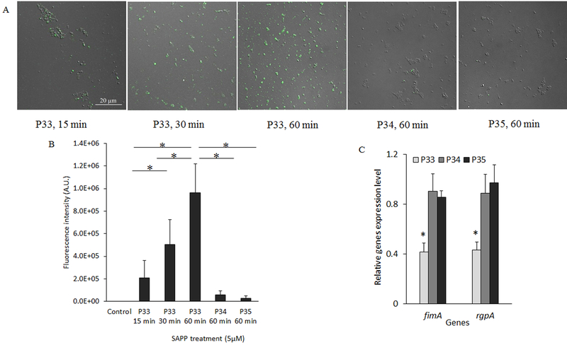
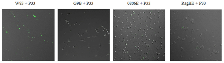
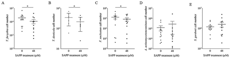
Porphyromonas gingivalis is a keystone bacterium in the oral microbial communities that elicits a dysbiosis between the microbiota and the host. Therefore, inhibition of this organism in dental plaques has been one of the strategies for preventing and treating chronic periodontitis. We previously identified a Streptococcal ArcA derived Anti-P gingivalils Peptide (SAPP) that in vitro, is capable of repressing the expression of several virulence genes in the organism. This leads to a significant reduction in P gingivalis virulence potential, including its ability to colonize on the surface of Streptococcus gordonii, to invade human oral epithelial cells, and to produce gingipains. In this study, we showed that SAPP had minimal cytotoxicity to human oral keratinocytes and gingival fibroblasts. We observed that SAPP directly bound to the cell surface of P gingivalis, and that alterations in the sequence at the N-terminus of SAPP diminished its abilities to interact with P gingivalis cells and repressed the expression of virulence genes. Most strikingly, we demonstrated using an ex-vivo assay that besides its inhibitory activity against P gingivalis colonization, SAPP could also reduce the levels of several other oral Gram-negative bacteria strongly associated with periodontitis in multispecies biofilms. Our results provide a platform for the development of SAPP-targeted therapeutics against chronic periodontitis.

Keywords: biofilms; oral microbiology; periodontal disease; porphyromonas.


Conflict of interest statement

We declare no conflict of interest.

Figures

Figure 1
Figure 1. Secondary structure analysis of SAPP by circular dichroism.
Far-UV CD spectrum of 0.75 μM SAPP acquired at room temperature in H2O. The spectrum shows that SAPP is disordered in solution with no significant population of helical or β-sheet secondary structure.
Figure 2
Figure 2. Determination of SAPP-induced cell membrane damage and cell death in HOKs and HPLFs.
HOKs and HPLFs were treated SAPP (0, 50, 200, and 800 μM) for 24 or 48 h. Cytotoxicity was determined by measuring LDH levels in the culture supernatant of HOKs and HPLFs (A and B). SAPP-induced cell death was quantitated using an Annexin V-FITC apoptosis kit (C and D). Each bar represents the mean ± standard deviation of LDH levels or numbers of apoptotic and necrotic cells. Data are the mean of at least three independent experiments.
Figure 3
Figure 3. Binding of fluorescence labeled SAPP to P. gingivalis.
(A) P. gingivalis 33277 biofilms were formed on saliva-coated glass and exposed to FITC-labeled SAPP (P33, 5 μM) for 0, 15, 30, and 60 min or its FITC-labeled derivatives (P34 and P35, 5 μM) for 1 h. Bacterial associated peptides were visualized using the confocal microscope. (B) Each bar represents average fluorescence intensity in 10 random areas (24 × 24 mm). Asterisks indicate significant difference in fluorescence intensities detected for samples exposed to the peptides, compared to that detected in the sample without exposure. (C) Each bar represents the relative expression level of fimA or rgpA/B in P. gingivalis 33277 grown with FITC-labeled SAPP (P33, 16 μM) or its derivatives (P34 and P35, 16 μM), which was compared to that in 33277 grown without SAPP (1 unit). Means and standard deviations were from three independent experiments. Asterisks indicate statistically significance between expression levels of genes in P. gingivalis 33277 grown with and without SAPP (control) (P < 0.05; t test).
Figure 4
Figure 4. Affinity of SAPP to bacterial surfaces.
The binding of SAPP to bacterial strains was assessed using confocal microscopy. FITC-labeled SAPP (P33, 5 μM) was incubated with P. gingivalis strainW83, S. gordonii G9B, or two P. gingivalis mutants (0806E and RagBE)for 1 h.
Figure 5
Figure 5. Attachment of P. gingivalis in multispecies biofilms.
(A) Multispecies biofilms were created using dental plaque samples retrieved from 11 chronic periodontitis patients, with sample from each patient divided into two cultures and incubated in the presence or absence of SAPP (48 μM). The level of P. gingivalis in the biofilms was determined using qPCR with P. gingivalis 16S rRNA primers. Mean and standard error of P. gingivalis cells detected in the biofilms are presented, and an asterisk indicates a significant difference between the cultures incubated with or without SAPP (p < 0.05; t test). (B) Comparison of P. gingivalis levels detected in the biofilms in the presence and absence of SAPP in each patient sample. Level of P. gingivalis in the biofilm without SAPP exposure is considered as 100%. The fimA genotype detected in each patient sample is indicated.
Figure 6
Figure 6. Determination of other oral bacterial levels in the biofilms generated using an ex-vivo assay.
Levels of T. forsythia (A), T. denticola (B), F. nucleatum (C), A. actinomycetemcomitans (D), and S. gordonii (E) in the biofilms were determined using qPCR with the corresponding specific 16S rRNA or 23S rRNA primers. Asterisks indicate statistical difference between two cultures of each sample grown with or without SAPP (P < 0.05; t−test).

References

    1. Kassebaum NJ, Bernabe E, Dahiya M, Bhandari B, Murray CJ, Marcenes W Global burden of severe periodontitis in 1990–2010: a systematic review and meta-regression. J Dent Res 2014;93:1045–1053. - PMC - PubMed
    1. Burt B, Research S, Therapy Committee of the American Academy of, P. Position paper: epidemiology of periodontal diseases. J Periodontol 2005;76:1406–1419. - PubMed
    1. Petersen PE, Ogawa H The global burden of periodontal disease: towards integration with chronic disease prevention and control. Periodontol 2000 2012;60:15–39. - PubMed
    1. Eke PI, Dye BA, Wei L, Thornton-Evans GO, Genco RJ, Cdc Periodontal Disease Surveillance workgroup: James Beck, GDRP. Prevalence of periodontitis in adults in the United States: 2009 and 2010. J Dent Res 2012;91:914–920. - PubMed
    1. Eke PI, Dye BA, Wei L et al. Update on Prevalence of Periodontitis in Adults in the United States: NHANES 2009 to 2012. J Periodontol 2015;86:611–622. - PMC - PubMed

Publication types

MeSH terms