Review
. 2018 Dec 14;64(6):469-476.
doi: 10.1262/jrd.2018-110. Epub 2018 Oct 8.

The roles of kisspeptin in the mechanism underlying reproductive functions in mammals

Affiliations
Review

The roles of kisspeptin in the mechanism underlying reproductive functions in mammals

Yoshihisa Uenoyama et al. J Reprod Dev. .

Abstract

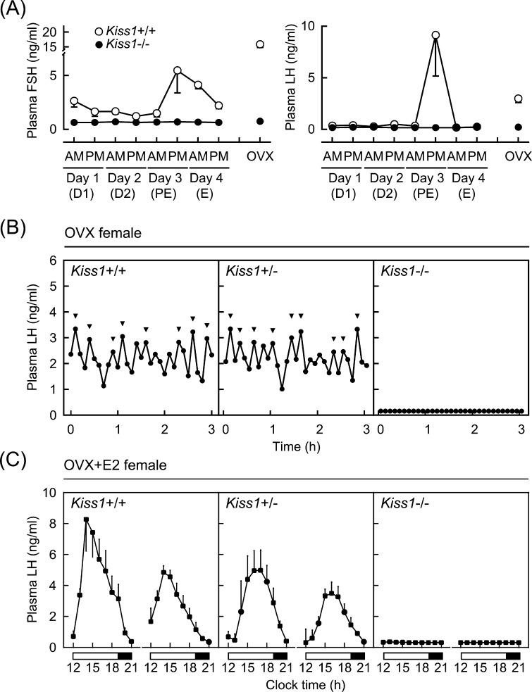
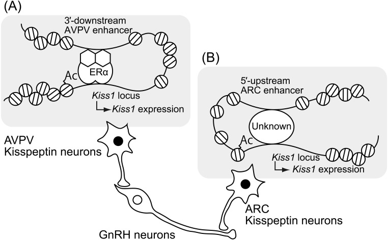
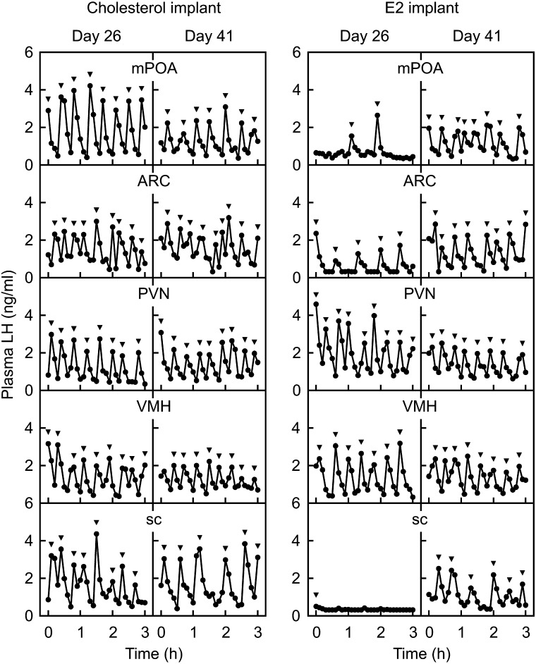
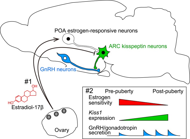
Kisspeptin, identified as a natural ligand of GPR54 in 2001, is now considered as a master regulator of puberty and subsequent reproductive functions in mammals. Our previous studies using Kiss1 knockout (KO) rats clearly demonstrated the indispensable role of kisspeptin in gonadotropin-releasing hormone (GnRH)/gonadotropin secretion. In addition, behavioral analyses of Kiss1 KO rats revealed an organizational effect of kisspeptin on neural circuits controlling sexual behaviors. Our studies using transgenic mice carrying a region-specific Kiss1 enhancer-driven reporter gene provided a clue as to the mechanism by which estrogen regulates Kiss1 expression in hypothalamic kisspeptin neurons. Analyses of Kiss1 expression and gonadotropin secretion during the pubertal transition shed light on the mechanism triggering GnRH/gonadotropin secretion at the onset of puberty in rats. Here, we summarize data obtained from the aforementioned studies and revisit the physiological roles of kisspeptin in the mechanism underlying reproductive functions in mammals.

Keywords: Estrogen; Gonadotropin-releasing hormone (GnRH); Kiss1; Luteinizing hormone (LH); Puberty.


Figures

Fig. 1.
Fig. 1.
Lack of pulsatile and surge modes of gonadotropin secretion in Kiss1 knockout (Kiss1–/–) rats. (A) Plasma follicle-stimulating hormone (FSH) and luteinizing hormone (LH) levels in wild-type (Kiss1+/+) and Kiss1–/– female rats. Plasma samples were collected from gonad-intact animals at 1000 h (AM) and 1700 h (PM) (1410 h light/dark cycle, light on 0500 h) for four consecutive days, and ovariectomized (OVX) rats at 1000 h. Stages of the estrous cycle were determined by vaginal smears in Kiss1+/+ rats. D1, diestrus 1; D2, diestrus 2; PE, proestrus; E, estrus. (B) Individual 3-h plasma LH profiles of representative OVX Kiss1+/+, heterozygous (Kiss1+/–), and Kiss1–/– rats. Arrowheads indicate LH pulses identified with the PULSAR computer program [70]. (C) Nine-hour plasma LH profiles of Kiss1+/+, Kiss1+/–, and Kiss1–/– female rats, which were OVX and received preovulatory levels of estradiol-17β (E2). Plasma samples were collected for two consecutive days. Open and closed horizontal bars indicate light and dark periods. Originally published in Uenoyama et al. (2015) [34].
Fig. 2.
Fig. 2.
Schematic illustrations of axonal projections of two populations of hypothalamic kisspeptin neurons and the molecular and epigenetic mechanism regulating Kiss1 expression in the anteroventral periventricular nucleus (AVPV) and arcuate nucleus (ARC). (A) Estrogen-ERα complex bound on the Kiss1 promoter region induces histone acetylation in the Kiss1 promoter region and forms chromatin loops between the 3′-downstream region and promoter region of the Kiss1 locus to activate AVPV Kiss1 expression. (B) Histone acetylation of the Kiss1 promoter region and chromatin loops formed between the 5′-upstream region and promoter region of the Kiss1 locus via unknown transcriptional factor(s) appeared to be involved in ARC Kiss1 expression in the absence of estrogen. Ac, histone acetylation.
Fig. 3.
Fig. 3.
Estrogen-dependent prepubertal suppression of pulsatile LH secretion in female rats. Plasma LH profiles in cholesterol- (left panel) or E2-implanted (right panel) OVX rats at 26 (prepubertal period) and 41 (postpubertal period) days of age. Note that E2 implanted into the medial preoptic area (mPOA), ARC or subcutaneous (sc) space, but not into the paraventricular nucleus (PVN) or ventromedial nucleus (VMH), suppressed LH pulses in OVX rats in the prepubertal period. Arrowheads indicate LH pulses identified with the PULSAR computer program [70]. Originally published in Uenoyama et al. (2015) [69].
Fig. 4.
Fig. 4.
Schematic illustration showing a possible mechanism regulating the pubertal augmentation of ARC Kiss1 expression to trigger pulsatile GnRH/gonadotropin secretion. #1, Estrogen strongly suppresses ARC Kiss1 expression via direct and indirect pathways: estrogen-responsive neurons in the POA mediate estrogen negative feedback action to ensure the prepubertal suppression of ARC Kiss1 expression. #2, During the pubertal transition, the sensitivity to the estrogen negative feedback action on ARC Kiss1 expression somehow decreases, which results in an increase in Kiss1 expression, and the subsequent secretion of kisspeptin triggers GnRH/gonadotropin secretion at pubertal onset.

References

    1. Schally AV, Arimura A, Kastin AJ, Matsuo H, Baba Y, Redding TW, Nair RM, Debeljuk L, White WF. Gonadotropin-releasing hormone: one polypeptide regulates secretion of luteinizing and follicle-stimulating hormones. Science 1971; 173: 1036–1038. - PubMed
    1. Amoss M, Burgus R, Blackwell R, Vale W, Fellows R, Guillemin R. Purification, amino acid composition and N-terminus of the hypothalamic luteinizing hormone releasing factor (LRF) of ovine origin. Biochem Biophys Res Commun 1971; 44: 205–210. - PubMed
    1. Silverman AJ, Antunes JL, Ferin M, Zimmerman EA. The distribution of luteinizing hormone-releasing hormone (LHRH) in the hypothalamus of the rhesus monkey. Light microscopic studies using immunoperoxidase technique. Endocrinology 1977; 101: 134–142. - PubMed
    1. Witkin JW, Paden CM, Silverman AJ. The luteinizing hormone-releasing hormone (LHRH) systems in the rat brain. Neuroendocrinology 1982; 35: 429–438. - PubMed
    1. Lehman MN, Robinson JE, Karsch FJ, Silverman AJ. Immunocytochemical localization of luteinizing hormone-releasing hormone (LHRH) pathways in the sheep brain during anestrus and the mid-luteal phase of the estrous cycle. J Comp Neurol 1986; 244: 19–35. - PubMed