. 2018 Oct 9;12(10):e0006815.
doi: 10.1371/journal.pntd.0006815. eCollection 2018 Oct.

Intrinsic activation of the vitamin D antimicrobial pathway by M. leprae infection is inhibited by type I IFN

Affiliations

Intrinsic activation of the vitamin D antimicrobial pathway by M. leprae infection is inhibited by type I IFN

Kathryn Zavala et al. PLoS Negl Trop Dis. .

Abstract

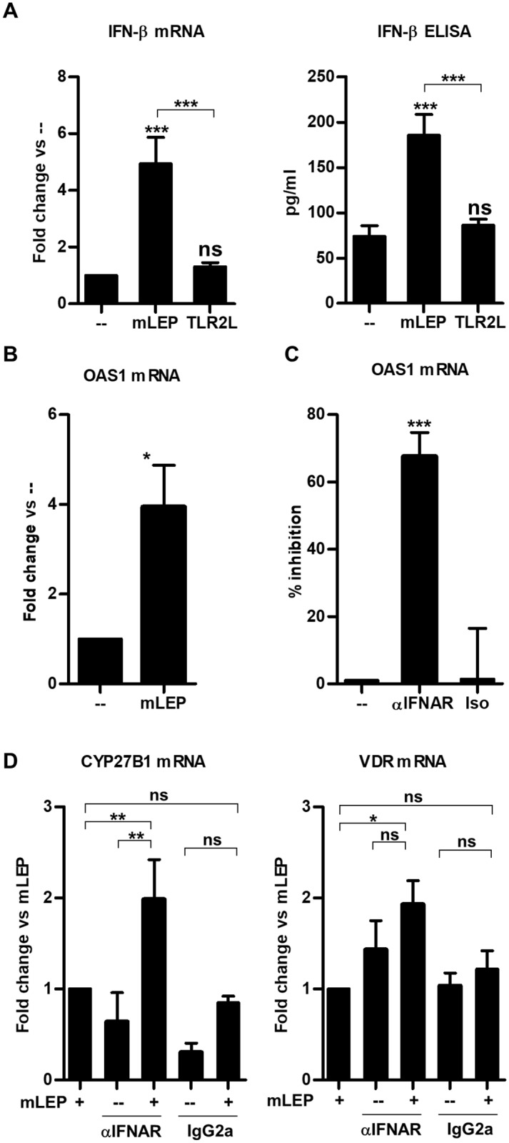
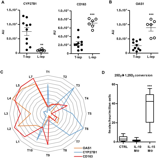
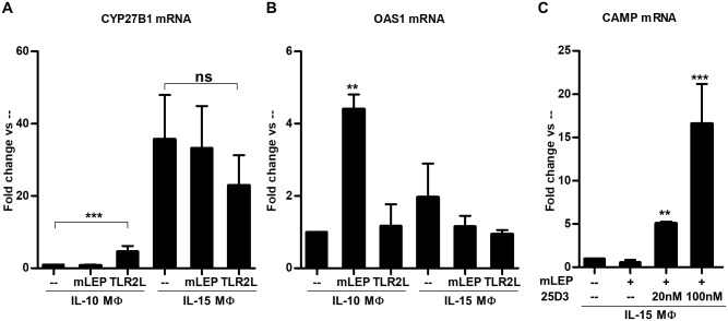
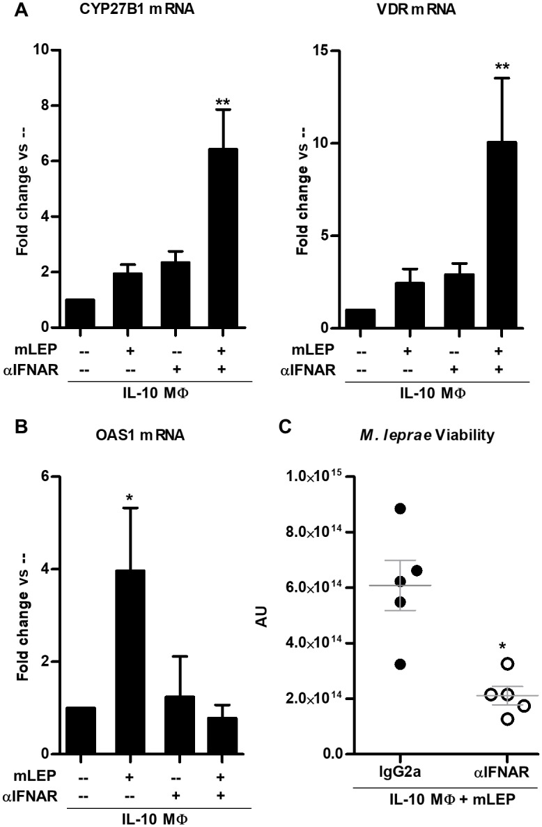
Following infection, virulent mycobacteria persist and grow within the macrophage, suggesting that the intrinsic activation of an innate antimicrobial response is subverted by the intracellular pathogen. For Mycobacterium leprae, the intracellular bacterium that causes leprosy, the addition of exogenous innate or adaptive immune ligands to the infected monocytes/macrophages was required to detect a vitamin D-dependent antimicrobial activity. We investigated whether there is an intrinsic immune response to M. leprae in macrophages that is inhibited by the pathogen. Upon infection of monocytes with M. leprae, there was no upregulation of CYP27B1 nor its enzymatic activity converting the inactive prohormone form of vitamin D (25-hydroxyvitamin D) to the bioactive form (1,25α-dihydroxyvitamin D). Given that M. leprae-induced type I interferon (IFN) inhibited monocyte activation, we blocked the type I IFN receptor (IFNAR), revealing the intrinsic capacity of monocytes to recognize M. leprae and upregulate CYP27B1. Consistent with these in vitro studies, an inverse relationship between expression of CYP27B1 vs. type I IFN downstream gene OAS1 was detected in leprosy patient lesions, leading us to study cytokine-derived macrophages (MΦ) to model cellular responses at the site of disease. Infection of IL-15-derived MΦ, similar to MΦ in lesions from the self-limited form of leprosy, with M. leprae did not inhibit induction of the vitamin D antimicrobial pathway. In contrast, infection of IL-10-derived MΦ, similar to MΦ in lesions from patients with the progressive form of leprosy, resulted in induction of type I IFN and suppression of the vitamin D directed pathway. Importantly, blockade of the type I IFN response in infected IL-10 MΦ decreased M. leprae viability. These results indicate that M. leprae evades the intrinsic capacity of human monocytes/MΦ to activate the vitamin D-mediated antimicrobial pathway via the induction of type I IFN.


Conflict of interest statement

The authors have declared that no competing interests exist.

Figures

Fig 1
Fig 1. CYP27B1 expression in leprosy and during M. leprae infection of primary human monocytes.
(A) One representative labeled section displaying CYP27b1 protein detection in leprosy lesions from one patient, (patient, n = 3), scale bar = 40 μm. Original magnification: x100. (B) Automated image analysis of CYP27b1 protein expression. Each dot represents the percentage of CYP27b1–stained area per nuclear area for each individual photomicrograph (n = 3 for each group). Data represent mean ± SEM. Statistical significance was determined using student t-test. Primary human monocytes were treated with M. leprae at a multiplicity of infection (MOI) of 10 or 100ng/mL Pam3CSK4 (TLR2L) for 24 hours and (C) CYP27B1 and gene expression levels were determined by quantitative PCR, or the cells were incubated with radiolabeled 25D3 for 5 hours, and (D) conversion to 1,25D3 was measured by high performance liquid chromatography (HPLC). Gene expression of (E) CYP24A1 and (F) conversion of 25D3 into 24,25D3 were also measured. Data are shown as mean ± SEM, n >3. Statistical significance was determined using one-way ANOVA. (* p ≤ 0.05, **p ≤ 0.01, ***p ≤ 0.001).
Fig 2
Fig 2. Blocking type I IFN signal during M. leprae infection relieves repression of intrinsic induction of CYP27B1.
(A) Primary human monocytes were infected with live M. leprae at MOI of 10 for 6 hours for mRNA and 24 hours for culture supernatants. Interferon beta 1 (IFN-β) expression was measured using quantitative PCR and protein levels measured using ELISA [17]. qPCR data are shown as mean fold change vs. media ± SEM, n = 5, and ELISA are shown as mean concentration ± SEM, n = 5. (B) Primary human monocytes were infected with M. leprae at MOI of 10 for 24 hours, and OAS1 gene expression was measured by qPCR. Data are shown as mean fold change vs. media ± SEM, n = 4. (C) Primary human monocytes were pre-treated with a monoclonal neutralizing antibody against the type I IFN receptor (IFNAR) or isotype control then infected with M. leprae for 24 hours. OAS1 gene expression was determined by qPCR and represented as percent inhibition. Data are shown as mean ± SEM, n = 8. (D) Monocytes were pretreated with IFNAR neutralizing antibody or isotype control then infected with M. leprae at MOI of 10 for 24 hours, then CYP27B1 and VDR gene expression levels were measured using quantitative PCR. Data are shown as mean ± SEM, n = 8. Statistical significance was determined using one-way ANOVA. (*p ≤ 0.05, **p ≤ 0.01, ***p ≤ 0.001).
Fig 3
Fig 3. CYP27B1 gene expression is negatively correlated with OAS1 gene expression in in vitro studies and in leprosy lesions.
(A) Correlation of CYP27B1 and CD163 as well as (B) OAS1 gene expression from microarray analysis of T-lep (n = 6) and L-lep (n = 5) human skin lesions. (C) Radar plot of CYP27B1, CD163 and OAS1 expression in T-lep and L-lep lesions normalized to the highest value in the experiment. (D) Primary human monocytes were treated with 10μg/mL IL-10 or 200ng/mL IL-15 for 48 hours. Control (Ctrl) MΦ were derived by treating monocytes with 103 U/mL IL-4 for 48 hours. IL-10 MΦ and IL-15 MΦ were incubated with radiolabeled 25D3 for 5 hours and conversion of 25D3 to 1,25D3 was measured by HPLC. Data are shown as mean ± SEM, n = 3. Statistical significance was determined using one-way ANOVA. (**p ≤ 0.01, ***p ≤ 0.001).
Fig 4
Fig 4. IL-15 MΦ and IL-10 MΦ differentially respond to M. leprae infection.
IL-15 MΦ and IL-10 MΦ were infected with M. leprae or stimulated with TLR2L for 24 hours. (A) CYP27B1 and (B) OAS1 gene expression levels were assayed by quantitative PCR. (C) IL-15 MΦ were infected with M. leprae and supplemented with 20nM 25D3, 100nM 25D3, or vehicle control for 24 hours. Cathelicidin (CAMP) gene expression was assayed by qPCR. Data are shown as mean fold change ± SEM, n = 3. Statistical significance was determined using one-way ANOVA. (*p ≤ 0.05, **p ≤ 0.01, ***p ≤ 0.001).
Fig 5
Fig 5. Blocking type I IFN signaling during M. leprae infection of IL-10 MΦ reduces bacterial viability.
IL-10 MΦ were pre-treated with either antibody against the type I IFN receptor, IFNAR, or isotype control then infected with M. leprae at an MOI of 10 for 24 hours. (A) CYP27B1, VDR, and (B) OAS1 gene expression levels were measured using qPCR. Data are shown as mean ± SEM, n < 5. (C) IL-10 MΦ were infected with M. leprae and co-treated with IFNAR neutralizing monoclonal antibody, or isotype control for 5 days under vitamin D-sufficient conditions. The ratio of M. leprae 16S RNA to RLEP DNA was calculated as a measurement of bacterial viability. Statistical significance was determined using one-way ANOVA. (*p ≤ 0.05, **p ≤ 0.01).

References

    1. Fabri M, Stenger S, Shin DM, Yuk JM, Liu PT, Realegeno S, et al. Vitamin D is required for IFN-gamma-mediated antimicrobial activity of human macrophages. Science translational medicine. 2011;3(104):104ra2 10.1126/scitranslmed.3003045 . - DOI - PMC - PubMed
    1. Liu PT, Stenger S, Li H, Wenzel L, Tan BH, Krutzik SR, et al. Toll-like receptor triggering of a vitamin D-mediated human antimicrobial response. Science. 2006;311(5768):1770–3. 10.1126/science.1123933 . - DOI - PubMed
    1. Rohde KH, Veiga DF, Caldwell S, Balazsi G, Russell DG. Linking the transcriptional profiles and the physiological states of Mycobacterium tuberculosis during an extended intracellular infection. PLoS pathogens. 2012;8(6):e1002769 10.1371/journal.ppat.1002769 . - DOI - PMC - PubMed
    1. Ridley DS. Histological classification and the immunological spectrum of leprosy. Bull World Health Organ. 1974;51(5):451–65. . - PMC - PubMed
    1. Montoya D, Cruz D, Teles RM, Lee DJ, Ochoa MT, Krutzik SR, et al. Divergence of macrophage phagocytic and antimicrobial programs in leprosy. Cell host & microbe. 2009;6(4):343–53. 10.1016/j.chom.2009.09.002 . - DOI - PMC - PubMed

Publication types

MeSH terms