

Buried Barrett's Esophagus with High-Grade Dysplasia after Radiofrequency Ablation
- PMID: 30300980
- PMCID: PMC6547340
- DOI: 10.5946/ce.2018.124
Buried Barrett's Esophagus with High-Grade Dysplasia after Radiofrequency Ablation
Abstract
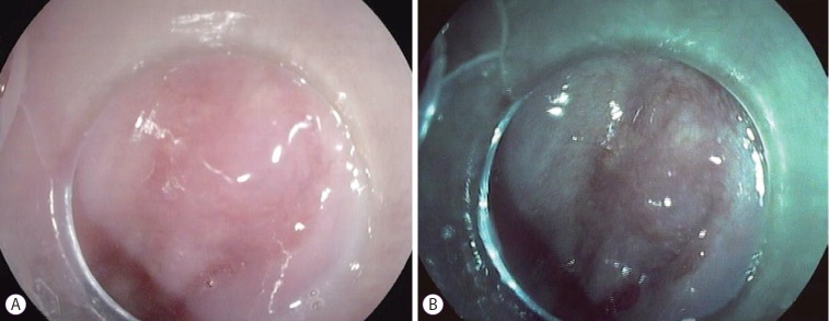
Radiofrequency ablation therapy is an effective endoscopic option for the eradication of Barrett's esophagus that appears to reduce the risk of esophageal cancer. A concern associated with this technique is the development of subsquamous/buried intestinal metaplasia, whose clinical relevance and malignant potential have not yet been fully elucidated. Fewer than 20 cases of subsquamous neoplasia after the successful radiofrequency ablation of Barrett's esophagus have been reported to date. Here, we describe a new case of subsquamous neoplasia (high-grade dysplasia) following radiofrequency ablation that was managed with endoscopic resection. Our experience suggests that a meticulous endoscopic inspection prior to and after radiofrequency ablation is fundamental to reduce the risk of buried neoplasia development.
Keywords: Barrett esophagus; Catheter ablation; Esophageal neoplasms.
Conflict of interest statement
Figures

Similar articles
-
Esophageal neoplasia arising from subsquamous buried glands after an apparently successful photodynamic therapy or radiofrequency ablation for Barrett's associated neoplasia.Scand J Gastroenterol. 2015;50(11):1315-21. doi: 10.3109/00365521.2015.1043578. Epub 2015 May 8. Scand J Gastroenterol. 2015. PMID: 25956748 Clinical Trial.
-
Recurrence of subsquamous dysplasia and carcinoma after successful endoscopic and radiofrequency ablation therapy for dysplastic Barrett's esophagus.Endoscopy. 2013 Jul;45(7):571-4. doi: 10.1055/s-0032-1326419. Epub 2013 Apr 16. Endoscopy. 2013. PMID: 23592390
-
Subsquamous intestinal metaplasia after ablation of Barrett's esophagus: frequency and importance.Curr Opin Gastroenterol. 2013 Jul;29(4):454-9. doi: 10.1097/MOG.0b013e3283622796. Curr Opin Gastroenterol. 2013. PMID: 23674187 Review.
-
Post-ablation buried neoplasia in Barrett's esophagus.Scand J Gastroenterol. 2021 May;56(5):624-628. doi: 10.1080/00365521.2021.1896774. Epub 2021 Mar 25. Scand J Gastroenterol. 2021. PMID: 33764825 Review.
-
AGA Clinical Practice Update on Endoscopic Treatment of Barrett's Esophagus With Dysplasia and/or Early Cancer: Expert Review.Gastroenterology. 2020 Feb;158(3):760-769. doi: 10.1053/j.gastro.2019.09.051. Epub 2019 Nov 12. Gastroenterology. 2020. PMID: 31730766 Review.
Cited by
-
Inflammatory bowel disease- and Barrett's esophagus-associated neoplasia: the old, the new, and the persistent struggles.Gastroenterol Rep (Oxf). 2019 Aug 13;7(6):379-395. doi: 10.1093/gastro/goz032. eCollection 2019 Dec. Gastroenterol Rep (Oxf). 2019. PMID: 31857901 Free PMC article. Review.
-
Management of Barrett Esophagus Following Radiofrequency Ablation.Gastroenterol Hepatol (N Y). 2019 Jul;15(7):377-386. Gastroenterol Hepatol (N Y). 2019. PMID: 31391808 Free PMC article.
-
Texture and Colour Enhancement Imaging versus White Light Endoscopy for Detection of Dysplasia within Barrett's Oesophagus: A Pilot Study.Digestion. 2025 Jun 16:1-9. doi: 10.1159/000546637. Online ahead of print. Digestion. 2025. PMID: 40523357 Free PMC article.
-
Indications, contraindications and limitations of endoscopic therapy for Barrett's esophagus and early esophageal adenocarcinoma.Therap Adv Gastroenterol. 2020 May 26;13:1756284820924209. doi: 10.1177/1756284820924209. eCollection 2020. Therap Adv Gastroenterol. 2020. PMID: 32523628 Free PMC article. Review.
References
-
- Falk GW. Barrett’s esophagus. Gastroenterology. 2002;122:1569–1591. - PubMed
-
- Simard EP, Ward EM, Siegel R, Jemal A. Cancers with increasing incidence trends in the United States: 1999 through 2008. CA Cancer J Clin. 2012;62:118–128. - PubMed
-
- Stein HJ, Siewert JR. Barrett’s esophagus: pathogenesis, epidemiology, functional abnormalities, malignant degeneration, and surgical management. Dysphagia. 1993;8:276–288. - PubMed
-
- Shaheen NJ, Sharma P, Overholt BF, et al. Radiofrequency ablation in Barrett’s esophagus with dysplasia. N Engl J Med. 2009;360:2277–2288. - PubMed
-
- Iyer PG. A “deeper” look at subsquamous structures beneath the neosquamous epithelium after Barrett’s esophagus endotherapy. Gastrointest Endosc. 2016;83:89–91. - PubMed
Publication types
LinkOut - more resources
Full Text Sources
