. 2018 Nov 21;38(47):10042-10056.
doi: 10.1523/JNEUROSCI.0015-18.2018. Epub 2018 Oct 9.

Unilateral, 3D Arm Movement Kinematics Are Encoded in Ipsilateral Human Cortex

Affiliations

Unilateral, 3D Arm Movement Kinematics Are Encoded in Ipsilateral Human Cortex

David T Bundy et al. J Neurosci. .

Abstract

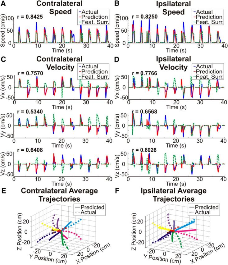
There is increasing evidence that the hemisphere ipsilateral to a moving limb plays a role in planning and executing movements. However, the exact relationship between cortical activity and ipsilateral limb movements is uncertain. We sought to determine whether 3D arm movement kinematics (speed, velocity, and position) could be decoded from cortical signals recorded from the hemisphere ipsilateral to the moving limb. By having invasively monitored patients perform unilateral reaches with each arm, we also compared the encoding of contralateral and ipsilateral limb kinematics from a single cortical hemisphere. In four motor-intact human patients (three male, one female) implanted with electrocorticography electrodes for localization of their epileptic foci, we decoded 3D movement kinematics of both arms with accuracies above chance. Surprisingly, the spatial and spectral encoding of contralateral and ipsilateral limb kinematics was similar, enabling cross-prediction of kinematics between arms. These results clarify our understanding that the ipsilateral hemisphere robustly contributes to motor execution and supports that the information of complex movements is more bihemispherically represented in humans than has been previously understood.SIGNIFICANCE STATEMENT Although limb movements are traditionally understood to be driven by the cortical hemisphere contralateral to a moving limb, movement-related neural activity has also been found in the ipsilateral hemisphere. This study provides the first demonstration that 3D arm movement kinematics can be decoded from human electrocorticographic signals ipsilateral to the moving limb. Surprisingly, the spatial and spectral encoding of contralateral and ipsilateral limb kinematics was similar. The finding that specific kinematics are encoded in the ipsilateral hemisphere demonstrates that the ipsilateral hemisphere contributes to the execution of unilateral limb movements, improving our understanding of motor control. Additionally, the bihemisheric representation of voluntary movements has implications for the development of neuroprosthetic systems for reaching and for neurorehabilitation strategies following cortical injuries.

Keywords: BCI; ECoG; electrocorticography; ipsilateral; reach.


Figures

Figure 1.
Figure 1.
Study methodology. Patients implanted with electrocorticography arrays completed a 3D center-out reaching task. A, Electrode locations were based upon the clinical requirements of each patient and were localized to an atlas brain for display. B, Patients were seated in the semirecumbent position and completed reaching movements from the center to the corners of a 50 cm physical cube based upon cues from LED lights located at each target while hand positions and ECoG signals were simultaneously recorded. Each patient was implanted with electrodes in a single cortical hemisphere and performed the task with the arm contralateral (C) and ipsilateral (D) to the electrode array in separate recording sessions. E, The task incorporated a center hold period (Hold-A), planning delay, movement period, and exterior hold period (Hold-B). To decode kinematics of contralateral and ipsilateral reaching movements, a hierarchical PLS regression that incorporated a logistic regression classification of movement and rest periods to switch the predicted output between the output of two PLS regression models was used. The first PLS model was trained using data from the rest periods to predict speed and velocity during rest periods and the second PLS regression model was trained using data from the movement periods to predict speed and velocity during movement periods (F). (E adapted from Bundy et al., 2016 under terms of the CC BY license).
Figure 2.
Figure 2.
Movement-related spectral power changes. A, After aligning to the movement onset, movement speed was averaged across trials and patients for the contralateral and ipsilateral hand showing similar amplitudes and time courses of reaching movements. The vertical lines indicate time windows for topographical plots of spectral power changes shown in B. B, Trial-averaged z-scores of log-transformed mu, beta, and high-gamma power changes that are significantly (p < 0.05) different from baseline are plotted for all electrodes at time windows 300 ms before movement onset (top), 100 ms before movement onset (middle), and 250 ms after movement onset (bottom). Movement-related decreases in mu and beta band power and movement-related increases in high-gamma band power are observed over sensorimotor cortex for both contralateral and ipsilateral reaches but begin earlier and are greater in amplitude for contralateral arm reaches. C, The difference in the timing of movement-related spectral power changes was quantified by calculating the percentage of electrodes with z-scores significantly (p < 0.05) different from 0 at each time window. Additionally, correlations between the time courses of the percentage of active electrodes during contralateral and ipsilateral arm movements were calculated at various time lags to determine the time lag with the peak correlation. Correlations >0.9 were observed for the mu, beta 1, and gamma 2 frequencies with peak time lags of 50 ms (contralateral leading), −100 ms (ipsilateral leading), and 50 ms (contralateral leading). Therefore, whereas there were slight differences in the timing of gross movement-related neural activity, these time differences were not consistent across frequencies. D, The difference in amplitude of movement-related spectral power changes was quantified by calculating the percentage of electrodes with significantly different spectral power changes between contralateral and ipsilateral arm movements. In mu, beta, and high-gamma bands, a subset of active electrodes demonstrate significantly (p < 0.05) greater amplitude spectral power changes during contralateral arm movements relative to ipsilateral arm movements.
Figure 3.
Figure 3.
Exemplar kinematic predictions. AD, Exemplar kinematic predictions of contralateral arm movement speed (A), ipsilateral arm movement speed (B), contralateral arm movement velocity (C), and ipsilateral arm movement velocity (D). Actual kinematic traces are shown in blue, predicted kinematic traces are shown in red, and an example surrogate prediction with reshuffled feature weights is shown in green. The plots were generated using consecutive trials from a single contralateral and ipsilateral test set from Patient 4. Kinematic predictions were made from 2 s before movement onset to the end of each trial and show accurate predictions of 3D kinematics for both the contralateral and ipsilateral arm. E, F, Movement trajectories were generated by concatenating predicted velocities within each trial, normalizing the trajectory times across trials, and averaging all trajectories for each target location. Averaged trajectories were generated using every test set from Patient 4 and end in the correct quadrant for each target for both contralateral and ipsilateral arm movements.
Figure 4.
Figure 4.
Prediction accuracy. Prediction accuracy was assessed by calculating the percentage of trajectories ending in the correct quadrant as well as calculating the correlation coefficient (Pearson's r) between the observed and predicted kinematics (speed, Vx, Vy, and Vz). Prediction accuracies were calculated for each of the 100 random test sets for each patient and chance accuracy was determined using two different surrogate datasets. Accuracies were combined across patients and randomly selected test sets. Boxes show the median, 25th percentile, and 75th percentile of accuracy. Whiskers show the range of accuracies with outliers >2.7 SDs indicated by a “+” symbol. Comparisons that are statistically significant (p < 0.05) after Bonferonni correction for the total number of comparisons are indicated by a “*” symbol. Across patients, the prediction accuracy and correlations between predicted and actual speed, Vx, Vy, and Vz were all significantly (p < 0.05) better than both surrogate distributions even after Bonferroni correction for the total number of true and cross-prediction comparisons. Furthermore, in addition to the comparisons across patients shown here, the prediction accuracies and correlations between the predicted and actually kinematics were also significantly better than surrogate predictions for each individual patient (see extended data Fig. 4-1, and Fig. 4-2).
Figure 5.
Figure 5.
Spatial, spectral, and temporal importance. The importance of individual locations, frequencies, and time lags for predicting kinematics was determined by converting prediction model weights to activation patterns for the logistic regression classifying movement and rest (movement classification) and movement-period PLS regression. A, Normalized activation pattern weights for the top 25% of weights are plotted on an atlas brain using a Gaussian kernel centered at each electrode site. Activation patterns across patients were combined onto a single atlas brain and areas with overlapping coverage were combined across patients using a weighted average based upon the distance from each electrode. Additionally, the normalized activation pattern weights were averaged across velocity components to produce plots for velocity. For each kinematic component, the most important cortical locations are centered over the central sulcus in primary sensorimotor cortex for both hands. B, Normalized activation pattern weights for the top 25% of weights across all electrodes, patients, and cross folds were combined for each feature type. Distributions of activation pattern weights are plotted with boxes showing the median, 25th percentile, and 75th percentile and whiskers showing the extent of weights. Outliers >2.7 SDs from the median are shown with a “+” symbol. Low frequencies including the beta band and LMP had the largest normalized activation patterns for movement classification, whereas movement kinematics (speed and velocity) were represented most strongly within LMP features followed by beta and high-gamma band features for both contralateral and ipsilateral arm movements. C, For channels and frequencies with activation pattern weights in the top 25% of weights, the time lag between neural activity and kinematics with the peak activation weight magnitude was determined. Histograms show the proportion of weights at each time lag. The logistic regression weights had a peak at a time lag of 0 s. For speed and velocity, the neural activity led the kinematics that were predicted with the majority of time lags falling between −500 ms and 0 s for both contralateral and ipsilateral arm movements.
Figure 6.
Figure 6.
Exemplar cross-prediction accuracy. AD, Exemplar kinematic cross-predictions were generated by using ipsilateral reaching movements to train our model and predict contralateral movement speed (A) and velocity (C) and using contralateral movements to train a model to predict ipsilateral arm speed (B) and velocity (D). Actual kinematic traces are shown in blue, predicted kinematics traces are shown in red, and an example surrogate prediction with reshuffled feature weights is shown in green. The plots were generated using consecutive test set trials from a single test set from Patient 4. Kinematics predictions were made from 2 s before movement onset to the end of each trial and show accurate predictions of 3D kinematics even when the prediction model was trained using reaching movements from the opposite hand.
Figure 7.
Figure 7.
Cross-prediction accuracy. Cross-prediction accuracy was assessed by calculating the percentage of trajectories ending in the correct quadrant as well as calculating the correlation coefficient (Pearson's r) between the observed and predicted kinematics (speed, Vx, Vy, and Vz). Accuracies were combined across patients and random test sets and the boxes show the median, 25th percentile, and 75th percentile of accuracy. Whiskers show the range of accuracies with outliers >2.7 SDs indicated by a “+” symbol. Comparisons that are statistically significant (p < 0.05) after Bonferonni correction for the total number of comparisons are indicated by a “*” symbol. Across patients, with the exception of the comparison between the 'ipsilateral training, contralateral testing' prediction and the temporal surrogate prediction for speed, the cross-prediction accuracy and correlations between predicted and actual speed, Vx, Vy, and Vz were all significantly (p < 0.05) better than either surrogate even after Bonferroni correction for the total number of comparisons, showing that some components of the ECoG representation of kinematics are conserved within a single cortical hemisphere for contralateral and ipsilateral arm movements.
Figure 8.
Figure 8.
Comparison of true and cross-prediction accuracies. True and cross-prediction accuracy were assessed by calculating the percentage of trajectories ending in the correct quadrant as well as calculating the correlation coefficient (Pearson's r) between the observed and predicted kinematics (speed, Vx, Vy, and Vz). Accuracies were combined across patients and random test sets and the boxes show the median, 25th percentile, and 75th percentile of accuracy. Whiskers show the range of accuracies with outliers >2.7 SDs indicated by a “+” symbol. Comparisons that are statistically significant (p < 0.05) after Bonferonni correction for the total number of comparisons are indicated by a “*” symbol. Across patients, whereas the accuracy and correlations between predicted and actual speed, Vx, Vy, and Vz were all significantly (p < 0.05) better than chance for both true and cross-predictions, true predictions were significantly (p < 0.05) better than the respective cross-predictions even after Bonferroni correction for the total number of comparisons.

References

    1. Aizawa H, Mushiake H, Inase M, Tanji J (1990) An output zone of the monkey primary motor cortex specialized for bilateral hand movement. Exp Brain Res 82:219–221. - PubMed
    1. Baskett JJ, Marshall HJ, Broad JB, Owen PH, Green G (1996) The good side after stroke: ipsilateral sensory-motor function needs careful assessment. Age Ageing 25:239–244. 10.1093/ageing/25.3.239 - DOI - PubMed
    1. Benjamini Y, Hochberg Y (1995) Controlling the false discovery rate: a practical and powerful approach to multiple testing. J Roy Stat Soc B Met 57:289–300.
    1. Benjamini Y, Yekutieli D (2001) The control of the false discovery rate in multiple testing under dependency. Ann Stat 29:1165–1188. 10.1214/aos/1013699998 - DOI
    1. Buch ER, Modir Shanechi A, Fourkas AD, Weber C, Birbaumer N, Cohen LG (2012) Parietofrontal integrity determines neural modulation associated with grasping imagery after stroke. Brain 135:596–614. 10.1093/brain/awr331 - DOI - PMC - PubMed

Publication types

LinkOut - more resources