. 2018 Nov 8;131(21):jcs213710.
doi: 10.1242/jcs.213710.

Rab34 small GTPase is required for Hedgehog signaling and an early step of ciliary vesicle formation in mouse

Affiliations

Rab34 small GTPase is required for Hedgehog signaling and an early step of ciliary vesicle formation in mouse

Shouying Xu et al. J Cell Sci. .

Abstract

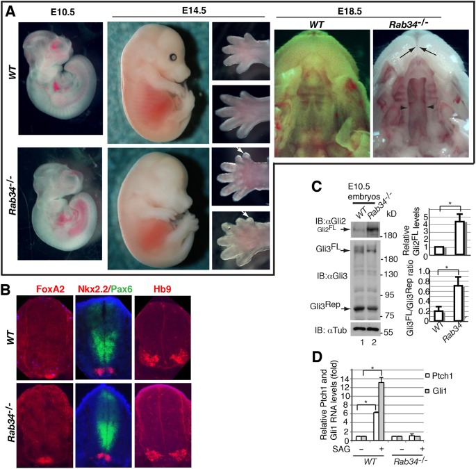
The primary cilium is a microtubule-based organelle that protrudes from the cell surface and plays essential roles in embryonic development. Ciliogenesis begins with the successive fusion of preciliary vesicles to form ciliary vesicles, which then dock onto the distal end of the mother centriole. Rab proteins have been linked to cilia formation in cultured cells, but not yet in vivo In the present study, we demonstrate that endocytic recycling protein Rab34 localizes to cilia, and that its mutation results in significant decrease of ciliogenesis in both cultured cells and mice. Rab34 is required for the successive fusion of preciliary vesicles to generate ciliary vesicles and for the migration of the mother centriole from perinuclear region to plasma membrane. We also show that Rab34 mutant mice exhibit polydactyly, and cleft-lip and -palate. These phenotypes are consistent with observations that nonciliated Rab34 mutant cells fail to respond to Hedgehog signaling and that processing of full-length Gli3 to its C-terminally truncated form is reduced in Rab34 mutant embryos. Therefore, Rab34 is required for an early step of ciliary vesicle formation and Hh signaling in vivo This article has an associated First Person interview with the first author of the paper.

Keywords: Cilia; Ciliary vesicle; Hedgehog; Rab34.


Conflict of interest statement

Competing interestsThe authors declare no competing or financial interests.

Figures

Fig. 1.
Fig. 1.
Rab34 is required for ciliogenesis in both cultured cells and in vivo. (A) Loss of Rab34 in cultured cells results in a significant decrease in ciliogenesis. NIH3T3 cells were stably infected with lentivirus that expressed sgRNAs as indicated. The cells were then immunostained for the ciliary marker Arl13b and counterstained with DAPI (nuclei). (Top) heterogeneous cell populations; (bottom) clonal cells, with clonal cells expressing FS-Rab34CR (right). The graph shows that the percentage of ciliated cells was significantly reduced in cells expressing Rab34 sgRNA1 or Rab34 sgRNA2, as compared to control GFP sgRNA. Ciliogenesis of clonal Rab34 sgRNA2 cells is rescued by overexpression of FS-Rab34CR. Two-tailed Student’s t-test P≤0.00028 (n=3 independent experiments, ≥100 cells counted for each category). (B) Loss of Rab34 results in a significant reduction in ciliogenesis in mouse tissues. Neural tube and limb bud sections at the forelimb area, and pMEFs of WT and Rab34 mutant were stained for the indicated ciliary markers. Graphs show the quantification of cilium numbers. Two-tailed Student’s t-test P≤0.000587 (n=5 neural tube sections from two different embryos were counted); *, significantly different.
Fig. 2.
Fig. 2.
Rab34 mutant mice exhibit polydactyly, cleft lip and palate, and reduced Hh signaling. (A) WT and Rab34 mutant embryos display polydactyly, and cleft lip and palate. For E14.5 embryos, arrows indicate extra digits. Lack of pigment in eyes of Rab34 mutant embryos is due to the segregation of albino from dark pigment, as heterozygous animals were maintained on a mixed albino SW and C57BL/6 background. For E18.5 embryos, arrows and arrowheads indicate cleft lip and palate, respectively. (B) Neural tube patterning is unaffected in Rab34 mutants. The neural tube sections around the forelimb areas of E10.5 embryos were stained for neuronal markers as indicated. All four markers appear to be specific, showing the normal pattern. For each genotype, ≥6 sections per marker and ≥3 embryos were used. (C) Gli3 processing is reduced in the Rab34 mutant. Western blot showing the levels of Gli2FL, Gli3FL, Gli3Rep and α-tubulin (a loading control). Quantification is shown in the bar graphs to the right. Two-tailed Student’s t-test P≤0.0115 (n=3 blots from 3 embryos). (D) Gli1 and Ptch1 RNA expression is not upregulated in Rab34 mutant pMEFs in response to stimulation with SAG. RT-qPCR shows relative RNA levels of Gli1 and Ptch1 in WT and Rab34 mutant pMEFs with or without stimulation with SAG. Two-tailed Student’s t-test P=0.00001 for both Gli1 and Ptch1 (n=3 independent experiments); *, significantly different.
Fig. 3.
Fig. 3.
Ciliated Rab34 mutant pMEFs and NIH3T3 cells are capable of responding to stimulation with the Smo agonist SAG. WT and Rab34 mutant pMEFs were incubated with vehicle or various concentrations of SAG overnight, and were then subjected to immunostaining of proteins as indicated above the panels (A) or to the left (B). Arrows indicate Gli2 staining in cilia. (C) Bar graph, showing quantification of data from three independent experiments. Note that both Smo and Gli2 accumulate in cilia of mutant cells upon stimulation with SAG. Two-tailed Student’s t-test P values between WT and mutant are 0.018, 0.235, 0.007 (Smo at 50, 100, 200 nM SAG) and 0.862, 0.001, 0.222 (Gli2 at 50, 100, 200 nM SAG) (n=3 independent experiments; in each category ≥100 cilia for WT and ≥10 cilia for mutant were counted). The large variation in the mutant is due to the small number of cilia being found. The overall trend is that that ciliated mutant cells show slightly weaker response to SAG stimulation compared to WT cells. (D) After overnight incubation with vehicle or SAG (200 nM), parental and Rab34 mutant NIH3T3 cells were stained for Smo and Arl13b. Note that Smo accumulated in cilia upon treatment with SAG in both parental and mutant cells. (E) The graph shows data from 3 independent experiments (≥130 cilia for WT and ≥11 cilia for mutant were counted for each experiment).
Fig. 4.
Fig. 4.
Rab34 localizes to cilia but its overexpression is not sufficient to promote ciliogenesis. (A) NIH3T3 cells stably expressing the indicated fusion proteins were stained for GFP, acetylated tubulin (ac-Tub) and DAPI (nucleus). Insets on right are enlargement (5x) of boxed area. (B) Graphs showing the quantitative staining results. Note that LAP-Rab34WT and LAP-Rab34Q111L (active mutant) but not LAP or LAP-Rab34T66N (dominant-negative inactive mutant), localized to cilia in >20% of cells that express either fusion protein (graph on left). However, overexpression of either LAP-Rab34WT or LAP-Rab34Q111L is not sufficient to induce ciliogenesis (graph on right), because two-tailed Student's t-test P≥0.0565 (n≥240 cells for each category). + indicates fusion proteins corresponding to columns. (C) Western blot showing expression of LAP and LAP-Rab34WT and mutants. α-Tubulin (αTub) was used as a loading control. Lane number corresponds to proteins in B.
Fig. 5.
Fig. 5.
Rab34 is not required for centrosomal and/or ciliary localization of Rab8, Rab11, Rabin8, BBS4, IFT20 and Cp110. WT NIH3T3 and Rab34 mutant NIH3T3 cells, and these cells overexpressing the indicated LAP fusion proteins were stained as indicated for GFP, endogenous proteins (IFT20 and Cp110), γ-tubulin (γTub) or acetylated and γ-tubulin (ac-+γTub) together. Ciliated cilia were selected for IFT20 and CP110, and a general population of cells was used for other markers. For each protein, except CP110, ≥10 images were taken. Note that the centriolar and ciliary localizations of all proteins examined are independent of Rab34. Graph on the right shows quantification of Cp110 staining at one centriole; two-tailed Student’s t-test P=0.816 (n≥54 cells). N.S., not significant.
Fig. 6.
Fig. 6.
Rab34 regulates the successive fusion of preciliary vesicles to form ciliary vesicles and migration of the basal bodies to cell cortex. (A) TEM micrographs of cilia on neuroepithelial cells in the neural tube near the forelimb area of E10.5 embryos of the indicated genotype (one embryo for each). Mother centrioles (MCs) are indicated by arrows, cilia are indicated by black arrowheads, representative preciliary vesicles are indicated by white arrowheads, ciliary vesicles are indicated by asterisks. The percentage and number of mother centrioles in each category are stated within each mircrograph; the total number of images (n) is given on the right of the panel. (B) The graph compares WT and mutant for each category. (C) Diagram showing the processes of cilia formation. Membrane structure is depicted in red. MC, mother centriole; DA, distal appendage; SDA, subdistal appendage; CV, cilia vesicle; PCV, periciliary vesicle; CB, cilia bud; CP, ciliary pocket; TZ, transition zone; AXN, axoneme; CM, cilia membrane; PM, plasma membrane.

References

    1. Bangs F. and Anderson K. V. (2017). Primary cilia and mammalian hedgehog signaling. Cold Spring Harb. Perspect Biol. 9 10.1101/cshperspect.a028175 - DOI - PMC - PubMed
    1. Boehlke C., Bashkurov M., Buescher A., Krick T., John A.-K., Nitschke R., Walz G. and Kuehn E. W. (2010). Differential role of Rab proteins in ciliary trafficking: Rab23 regulates smoothened levels. J. Cell Sci. 123, 1460-1467. 10.1242/jcs.058883 - DOI - PubMed
    1. Briscoe J., Pierani A., Jessell T. M. and Ericson J. (2000). A homeodomain protein code specifies progenitor cell identity and neuronal fate in the ventral neural tube. Cell 101, 435-445. 10.1016/S0092-8674(00)80853-3 - DOI - PubMed
    1. Campeau E., Ruhl V. E., Rodier F., Smith C. L., Rahmberg B. L., Fuss J. O., Campisi J., Yaswen P., Cooper P. K. and Kaufman P. D. (2009). A versatile viral system for expression and depletion of proteins in mammalian cells. PLoS ONE 4, e6529 10.1371/journal.pone.0006529 - DOI - PMC - PubMed
    1. Chen J. K., Taipale J., Young K. E., Maiti T. and Beachy P. A. (2002). Small molecule modulation of Smoothened activity. Proc. Natl. Acad. Sci. USA 99, 14071-14076. 10.1073/pnas.182542899 - DOI - PMC - PubMed

Publication types