. 2018 Oct 9;9(5):e01738-18.
doi: 10.1128/mBio.01738-18.

Inducible Nitric Oxide Synthase Is a Key Host Factor for Toxoplasma GRA15-Dependent Disruption of the Gamma Interferon-Induced Antiparasitic Human Response

Affiliations

Inducible Nitric Oxide Synthase Is a Key Host Factor for Toxoplasma GRA15-Dependent Disruption of the Gamma Interferon-Induced Antiparasitic Human Response

Hironori Bando et al. mBio. .

Abstract

Although Toxoplasma virulence mechanisms targeting gamma interferon (IFN-γ)-induced cell-autonomous antiparasitic immunity have been extensively characterized in mice, the virulence mechanisms in humans remain uncertain, partly because cell-autonomous immune responses against Toxoplasma differ markedly between mice and humans. Despite the identification of inducible nitric oxide synthase (iNOS) as an anti-Toxoplasma host factor in mice, here we show that iNOS in humans is a pro-Toxoplasma host factor that promotes the growth of the parasite. The GRA15 Toxoplasma effector-dependent disarmament of IFN-γ-induced parasite growth inhibition was evident when parasite-infected monocytes were cocultured with hepatocytes. Interleukin-1β (IL-1β), produced from monocytes in a manner dependent on GRA15 and the host's NLRP3 inflammasome, combined with IFN-γ to strongly stimulate iNOS expression in hepatocytes; this dramatically reduced the levels of indole 2,3-dioxygenase 1 (IDO1), a critically important IFN-γ-inducible anti-Toxoplasma protein in humans, thus allowing parasite growth. Taking the data together, Toxoplasma utilizes human iNOS to antagonize IFN-γ-induced IDO1-mediated cell-autonomous immunity via its GRA15 virulence factor.IMPORTANCEToxoplasma, an important intracellular parasite of humans and animals, causes life-threatening toxoplasmosis in immunocompromised individuals. Gamma interferon (IFN-γ) is produced in the host to inhibit the proliferation of this parasite and eventually cause its death. Unlike mouse disease models, which involve well-characterized virulence strategies that are used by Toxoplasma to suppress IFN-γ-dependent immunity, the strategies used by Toxoplasma in humans remain unclear. Here, we show that GRA15, a Toxoplasma effector protein, suppresses the IFN-γ-induced indole-2,3-dioxygenase 1-dependent antiparasite immune response in human cells. Because NLRP3-dependent production of IL-1β and nitric oxide (NO) in Toxoplasma-infected human cells is involved in the GRA15-dependent virulence mechanism, blocking NO or IL-1β production in the host could represent a novel therapeutic approach for treating human toxoplasmosis.

Keywords: Toxoplasma gondii; cell-autonomous immunity; host-parasite interaction; human immunology; immune suppression; interferon.


Figures

FIG 1
FIG 1
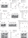
GRA15 suppresses IFN-γ-induced parasite reduction under THP-1/Huh7 coculture conditions. (A) THP-1 or Huh7 cells were left untreated (medium only [Med]) or treated with IFN-γ for 24 h and then infected with Pru T. gondii. The parasite survival rates were measured by luciferase assay. (B) THP-1 cells were infected with WT or GRA15-KO Pru T. gondii. The infected THP-1 cells were cocultured with Huh7 cells and then left untreated or treated with IFN-γ. The parasite survival rates were measured by luciferase assay. (C) THP-1 cells were infected with GRA15-KO Pru T. gondii stably expressing GRA15 or empty vectors. The infected THP-1 cells were cocultured with Huh7 cells and then left untreated or treated with IFN-γ. The parasite survival rates were measured by luciferase assay. (D) THP-1 or Huh7 cells were left untreated or treated with IFN-γ for 24 h and then infected with wild-type or GRA15-KO Pru T. gondii. The parasite numbers were calculated by quantification of the amount of genomic DNA of the sag1 gene, which was measured by qPCR, using the standard curve shown in Fig. S1F. (E) THP-1 cells were infected with WT or GRA15-KO Pru T. gondii. The infected THP-1 cells were cocultured with Huh7 cells and then left untreated or treated with IFN-γ. The numbers were calculated by quantification of the amount of genomic DNA of the sag1 gene, which was measured by qPCR, using the standard curve shown in Fig. S1F. (F) BMDMs were infected with WT or GRA15-KO Pru T. gondii. The infected BMDMs were cocultured with primary mouse hepatocytes and then left untreated or treated with IFN-γ. The parasite survival rates were measured by luciferase assay. Indicated values represent means ± standard deviations (SD) (three biological replicates per group from three independent experiments) (panels A to F). *, P < 0.05; N.S., not significant (Student's t test).
FIG 2
FIG 2
IL-1β production and signaling are important for the GRA15-dependent virulence mechanism. (A) THP-1 cells were infected with WT or GRA15-KO Pru T. gondii. The filtered culture supernatant was placed directly on top of the Huh7 cells, and the cells were newly infected with WT or GRA15-KO Pru T. gondii and then left untreated or treated with IFN-γ. The parasite survival rates were measured by luciferase assay. (B) THP-1 cells were left uninfected or infected with WT or GRA15-KO Pru T. gondii (left) or with GRA15-KO Pru T. gondii stably expressing GRA15 or empty vectors (right). Levels of IL-1β released into the culture supernatant were measured by ELISA. (C) WT, MyD88-KO, or IL-1R1-KO Huh7 cells were left untreated or treated with the indicated cytokines for 24 h and then infected with Pru T. gondii. The parasite survival rates were measured by luciferase assay. (D) THP-1 cells were infected with Pru T. gondii. The infected THP-1 cells were cocultured with WT, MyD88-KO, or IL-1R1-KO Huh7 cells and then left untreated or treated with IFN-γ. The parasite survival rates were measured by luciferase assay. Indicated values represent means ± SD (three biological replicates per group from three independent experiments) (panels A to D). **, P < 0.01; * P < 0.05; N.S., not significant (Student's t test).
FIG 3
FIG 3
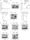
Caspase-1 and NLRP3 in THP-1 cells are involved in T. gondii-mediated IDO1 inhibition. (A and B) WT, CASP1-KO, NLRP1-KO, or NLRP3-KO THP-1 cells were left uninfected or infected with Pru T. gondii. (A) Levels of IL-1β released into the culture supernatant were measured by ELISA. (B) The infected THP-1 cells were cocultured with Huh7 cells and then left untreated or treated with IFN-γ. The parasite survival rates were measured by luciferase assay. Indicated values represent means ± SD (three biological replicates per group from three independent experiments) (panels A and B). ***, P < 0.001; **, P < 0.01; N.S., not significant (Student's t test).
FIG 4
FIG 4
IL-1β production and signaling are essential for GRA15-mediated IDO1 inhibition. (A) WT or MyD88-KO Huh7 cells were left untreated or treated with the indicated cytokines. Expression of IDO1 in the cell lysates was detected by Western blotting. (B and C) WT, Atg16L1-KO, or IDO1-KO Huh7 cells were left untreated or treated with IFN-γ for 24 h and then infected with Pru T. gondii. (B) The parasite survival rates were measured by luciferase assay. (C) The parasite number per vacuole after 24 h postinfection was measured by indirect immunofluorescent assay (IFA). (D) WT or IDO1-KO Huh7 cells were left untreated or treated with the indicated cytokines for 24 h and then infected with Pru T. gondii. The parasite survival rates were measured by luciferase assay. (E) THP-1 cells were infected with WT or GRA15-KO Pru T. gondii. The infected THP-1 cells were cocultured with Huh7 cells and then left untreated or treated with IFN-γ. Expression of IDO1 in the cell lysates was detected by Western blotting. (F) THP-1 cells were infected with GRA15-KO Pru T. gondii. The infected THP-1 cells were cocultured with WT or IDO1-KO Huh7 cells and then left untreated or treated with IFN-γ. The parasite survival rates were measured by luciferase assay. (G) THP-1 cells were infected with Pru T. gondii. The infected THP-1 cells were cocultured with WT or MyD88-KO Huh7 cells and then left untreated or treated with IFN-γ. Expression of IDO1 in cell lysates was detected by Western blotting. (H) WT, CASP1-KO, NLRP1-KO, or NLRP3-KO THP-1 cells were left uninfected or infected with Pru T. gondii. The infected THP-1 cells were cocultured with Huh7 cells and then left untreated or treated with IFN-γ. Expression of IDO1 in the cell lysates was detected by Western blotting. (I) WT or Irgm1/3 double-knockout (DKO) primary mouse hepatocytes were left untreated or treated with the indicated cytokines for 24 h and then infected with Pru T. gondii. The parasite survival rates were measured by luciferase assay. (J) Primary human hepatocyte or primary mouse hepatocytes were left untreated or treated with the indicated cytokines. Expression of indicated proteins in cell lysates was detected by Western blotting. (K) WT or IRGM-KO Huh7 cells were left untreated or treated with the indicated cytokines for 24 h and then infected with wild-type Pru T. gondii. The parasite survival rates were measured by luciferase assay. (L) WT or IRGM-KO Huh7 cells were left untreated or treated with the indicated cytokines. Expression of indicated proteins in cell lysates was detected by Western blotting. Each Western blot image is representative of three independent experiments (A, E, G, H, J, and L). Indicated values represent means ± SD (three biological replicates per group from three independent experiments) (B to D, F, I, and K). **, P < 0.01; N.S., not significant (Student's t test).
FIG 5
FIG 5
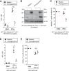
iNOS expression in Huh7 cells is required for GRA15-mediated IDO1 inhibition. (A) Quantitative RT-PCR analysis of iNOS mRNA level in WT or MyD88-KO Huh7 cells that were left untreated or treated with the indicated cytokines was performed. (B) WT or iNOS-KO Huh7 cells were left untreated or treated with the indicated cytokines. Expression of indicated proteins in the cell lysates was detected by Western blotting. (C) WT, iNOS-KO, or IDO1-KO Huh7 cells were left untreated or treated with the indicated cytokines for 24 h and then infected with Pru T. gondii. The parasite survival rates were measured by luciferase assay. (D to F) THP-1 cells were infected with Pru T. gondii. The infected THP-1 cells were cocultured with WT or iNOS-KO Huh7 cells and then left untreated or treated with IFN-γ. (D) Levels of NO2 released into the culture supernatant were measured by ELISA. (E) Expression of indicated proteins in the cell lysates was detected by Western blotting. (F) The parasite survival rates were measured by luciferase assay. (G to K) WT or CASP1-KO (G) THP-1 cells were infected with WT or GRA15-KO Pru T. gondii (H) or with GRA15-KO parasites stably expressing empty or GRA15 expression vectors (K). The infected THP-1 cells were cocultured with WT, MyD88-KO (I), or IL-1R1-KO (J) Huh7 cells and then left untreated or treated with IFN-γ. Expression of indicated proteins in the cell lysates was detected by Western blotting. Each Western blot image is representative of three independent experiments (B, E, and G to K). Indicated values represent means ± SD (three biological replicates per group from three independent experiments) (A, C, D, and F). **, P < 0.01; N.S., not significant (Student's t test).
FIG 6
FIG 6
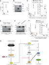
iNOS inhibitor prevents GRA15-dependent production of NO and reduction of IDO1 proteins. (A to C) THP-1 cells were infected with Pru T. gondii. The infected THP-1 cells were cocultured with Huh7 cells and then left untreated or treated with IFN-γ and/or aminoguanidine. (A) The levels of NO2 released into the culture supernatant were measured by ELISA. (B) Expression of indicated proteins in the cell lysates was detected by Western blotting. (C) The parasite survival rates were measured by luciferase assay. (D and E) WT or IDO1-KO Huh7 cells were left untreated or treated with the indicated cytokines and/or aminoguanidine and infected with Pru T. gondii. (D) Levels of NO2 released into the culture supernatant were measured by ELISA. (E) The parasite survival rates were measured by luciferase assay. The Western blot image is representative of three independent experiments (B). Indicated values represent means ± SD (three biological replicates per group from three independent experiments) (A, C, D, and E). ***, P < 0.001; **, P < 0.01; N.S., not significant (Student's t test).
FIG 7
FIG 7
The GRA15-dependent virulence mechanism is operative under primary human monocyte/hepatocyte coculture conditions. (A) Primary human monocytes were left uninfected or infected with WT or GRA15-KO Pru T. gondii. Levels of IL-1β released into the culture supernatant were measured by ELISA. (B) Primary human hepatocytes were left untreated or treated with the indicated cytokines. Expression of indicated proteins in the cell lysates was detected by Western blotting. (C and D) Primary human monocytes were infected with WT or GRA15-KO Pru T. gondii. The infected monocytes were cocultured with primary human hepatocytes and then left untreated or treated with IFN-γ. (C, left panel) Levels of NO2 released into the culture supernatant were measured by ELISA. (C, right panel) The parasite survival rates were measured by luciferase assay. (D) Expression of the indicated proteins in the cell lysates was detected by Western blotting. (E and F) Primary human monocytes were infected with Pru T. gondii. The infected monocytes were cocultured with primary human hepatocytes and then left untreated or treated with IFN-γ and/or aminoguanidine. (E) Expression of indicated proteins in the cell lysates was detected by Western blotting. (F, left panel) NO2 released into the culture supernatant was measured by ELISA. (F, right panel) The parasite survival rates were measured by luciferase assay. (G) GRA15-mediated indirect virulence program targeting IDO1 in the coculture model. T. gondii secreted effector GRA15 in the infected monocytes, which resulted in NLRP3-dependent IL-1β production. IL-1β from infected monocytes and IFN-γ induce the expression of iNOS and NO production, leading to indirect reduction of levels of IDO1 proteins in hepatocytes and allowing T. gondii growth. Each Western blot image is representative of three independent experiments (B, D, and E). Indicated values represent means ± SD (three biological replicates per group from three independent experiments) (A, C, and F). **, P < 0.01; N.S., not significant (Student's t test).

Similar articles

Cited by

References

    1. Dubey JP. 2010. Toxoplasmosis of animals and humans. CRC Press, Boca Raton, FL.
    1. Boothroyd JC. 2009. Toxoplasma gondii: 25 years and 25 major advances for the field. Int J Parasitol 39:935–946. doi:10.1016/j.ijpara.2009.02.003. - DOI - PMC - PubMed
    1. Montoya JG, Remington JS. 2008. Management of Toxoplasma gondii infection during pregnancy. Clin Infect Dis 47:554–566. doi:10.1086/590149. - DOI - PubMed
    1. Weitberg AB, Alper JC, Diamond I, Fligiel Z. 1979. Acute granulomatous hepatitis in the course of acquired toxoplasmosis. N Engl J Med 300:1093–1096. doi:10.1056/NEJM197905103001907. - DOI - PubMed
    1. Frenkel JK, Remington JS. 1980. Hepatitis in toxoplasmosis. N Engl J Med 302:178–179. - PubMed

Publication types

MeSH terms