. 2018 Oct 9;9(1):4179.
doi: 10.1038/s41467-018-06724-x.

Cryo-EM structure of the hibernating Thermus thermophilus 100S ribosome reveals a protein-mediated dimerization mechanism

Affiliations

Cryo-EM structure of the hibernating Thermus thermophilus 100S ribosome reveals a protein-mediated dimerization mechanism

Rasmus Kock Flygaard et al. Nat Commun. .

Abstract

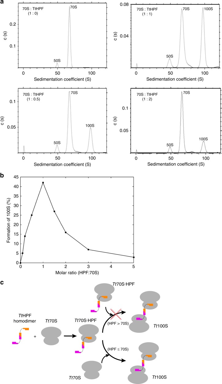
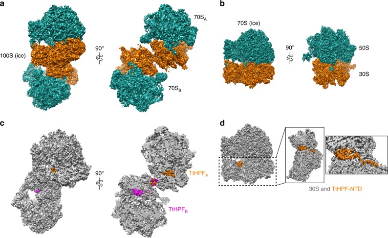
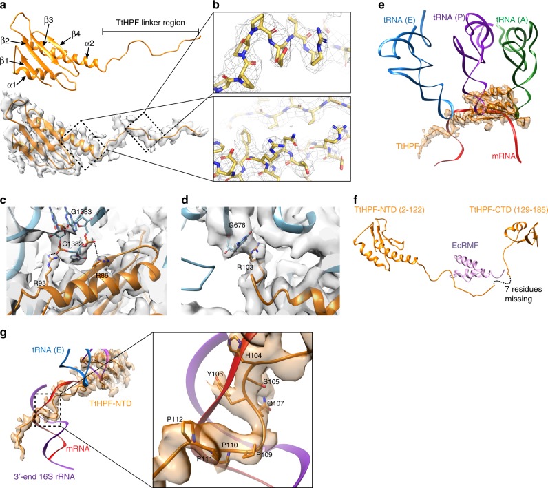
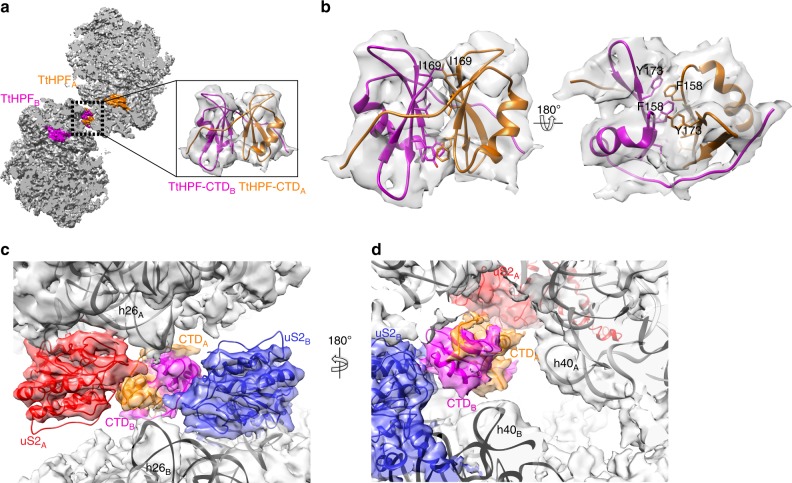
In response to cellular stresses bacteria conserve energy by dimerization of ribosomes into inactive hibernating 100S ribosome particles. Ribosome dimerization in Thermus thermophilus is facilitated by hibernation-promoting factor (TtHPF). In this study we demonstrate high sensitivity of Tt100S formation to the levels of TtHPF and show that a 1:1 ratio leads to optimal dimerization. We report structures of the T. thermophilus 100S ribosome determined by cryo-electron microscopy to average resolutions of 4.13 Å and 4.57 Å. In addition, we present a 3.28 Å high-resolution cryo-EM reconstruction of a 70S ribosome from a hibernating ribosome dimer and reveal a role for the linker region connecting the TtHPF N- and C-terminal domains in translation inhibition by preventing Shine-Dalgarno duplex formation. Our work demonstrates that species-specific differences in the dimerization interface govern the overall conformation of the 100S ribosome particle and that for Thermus thermophilus no ribosome-ribosome interactions are involved in the interface.


Conflict of interest statement

The authors declare no competing interests.

Figures

Fig. 1
Fig. 1
Analysis of in vitro TtHPF dependent formation of Tt100S. a Analytical ultracentrifugation sedimentation profiles show 70S ribosome as control (upper left), 70S ribosome mixed with TtHPF in 0.5 times molar ratio (lower left), 70S ribosome mixed with TtHPF in equimolar ratio (upper right) and 70S ribosome mixed with TtHPF in two times molar ratio (lower right). Formation of Tt100S ribosome is evident by the peak at a sedimentation coefficient of 100S. b Graphical representation of Tt100S ribosome formation from all AUC experiments. Formation of Tt100S ribosome by TtHPF is maximal in the case where the molar ratio of TtHPF to Tt70S is 1:1. See also Supplementary Figure 3. c Schematic illustration of TtHPF and Tt70S binding events leading to Tt100S ribosome formation. Binding of one NTD of TtHPF homodimer to Tt70S leads to a complex of Tt70S·TtHPF. In the case of sub- or equimolar ratios of TtHPF and Tt70S, binding of a vacant Tt70S ribosome to the free NTD of the Tt70S·TtHPF complex leads to Tt100S formation. However, in the case of TtHPF being present in excess molar ratios, Tt100S ribosome formation is inhibited because all Tt70S ribosomes bind a TtHPF homodimer
Fig. 2
Fig. 2
Cryo-EM structures of 100S (ice) and 70S (ice). a Orthogonal views of 100S (ice) with 50S subunits in green and 30S subunits in orange showing the two 70S copies constituting the 100S particle. b Orthogonal views of 70S (ice), coloring of subunits as in (a). c Views of 100S (ice) and slice-through view with both 70S ribosome copies colored in gray and the two TtHPF protein molecules colored in orange and magenta showing location of TtHPF-NTD and CTD within the 100S ribosome dimer. d View of 70S (ice) with TtHPF-NTD colored in orange. Close-up views on 30S subunit show location of TtHPF-NTD and the linker region. There was no density for the TtHPF-CTD in 70S (ice) reconstruction
Fig. 3
Fig. 3
Structure of TtHPF-NTD and its interactions with the 30S subunit. a Structure of TtHPF-NTD shown in orange cartoon. The corresponding cryo-EM density of 70S (ice), shown as semi-transparent gray surface, shows clear density for linker region. Secondary structure elements are labeled. b Close-up view on the high quality density map of the TtHPF-NTD (upper) and linker (lower) with model inside. Side chains are clearly resolved in the high-resolution density. c Close-up view on electrostatic interactions between TtHPF-NTD and ribosome centered around Arg86 and Arg93 interacting with phosphate backbone on nucleotides C1382 and G1383 in h44. d Example of stacking interaction between TtHPF-NTD residue Arg103 and nucleotide G676 of h23 in 16S rRNA. e Structure of TtHPF-NTD and its density (orange) superimposed with A-, P-, and E-site tRNAs (green, purple, blue) and mRNA (red) bound in 70S ribosome (PDB entry 4V6F). The binding position of TtHPF-NTD on the 30S subunit clearly overlaps with binding sites for all three tRNAs as well as mRNA. f Structure of TtHPF with the missing seven residues of the linker indicated by dashed line. The structure is superimposed with the chimeric structure of Tt70S-RMF (PDB entry 4V8G) showing the location of the E. coli RMF protein (violet) closely matching the binding position of the TtHPF linker region. g Coloring as in E now with 3′ end of 16S rRNA shown in violet. The linker region of TtHPF occupies a binding position on the 30S subunit that overlaps with the helix formed by mRNA and 3′ end of 16S rRNA. Close-up view shows TtHPF linker region residues His104, Ser105, Tyr106, and Gln107 overlapping with Shine–Dalgarno duplex between mRNA and 3′ end of 16S rRNA. Proline residues 109–112 overlap with mRNA binding position as well
Fig. 4
Fig. 4
Cryo-EM structure of 100S (amc). a Slice-through view of 100S (amc) reconstruction with both 70S ribosome copies colored in gray and the two TtHPF protein molecules colored in orange and magenta showing location of TtHPF-NTD and CTD within the 100S ribosome dimer. Close-up view shows the homodimer TtHPF-CTD model in the corresponding EM density. b Interactions between the two copies of TtHPF-CTD are governed by hydrophobic interactions (e.g., by Ile169) between the two beta-sheets (left side view) and by stacking interactions of aromatic residues (right side view). c Tt100S ribosome (light gray semi-transparent surface) dimerization interface around TtHPF-CTD (orange and magenta) with uS2 (red and blue models and densities) and h26 of 16S rRNA (labeled model inside 100S (amc) density). The length of h26 is too short for making interactions with uS2 on the other ribosome copy. d Coloring is as in C. h40 16S rRNA model is shown. There is no interaction from TtHPF-CTD homodimer to either of the h40 on the ribosomes
Fig. 5
Fig. 5
Conformation of Tt100S compared with other 100S structures. a Location of interacting proteins and rRNA as suggested by Polikanov et al. are shown in colors, uS2 in blue, uS3 in green, uS4 in purple, uS5 in magenta, uS9 in orange, uS10 in salmon, and h39 in gold. Tt100S ribosome density is colored gray. Clearly, the proteins and h39 are located far from each other. b B. subtilis 100S (EMD-3664, green) and S. aureus 100S (EMD-3637, purple and EMD-3638, salmon) in orthogonal views showing high degree of conformational conservation between the 100S molecules. c Same colors as in B. Tt100S (ice) shown in gray and superimposed to B. subtilis 100S. The orthogonal views clearly show are different conformation of the two ribosomes in Tt100S. d Tt100S (ice) in gray compared with E. coli 100S reconstructions (EMD-5174, pink and EMD-1750, light blue). The staggered conformation of Tt100S is very different from the E. coli 100S conformation, which is more back-to-back for the 30S subunits
Fig. 6
Fig. 6
Comparison of Tt100S ribosome to complexes of 30S-RNAP and 50S-HflX. a Structure of 30S-RNAP (PDB entry 6AWB) with 30S subunit in blue, RNAP in pink and RNAP β′ subunit in purple fitted in Tt100S (ice) density. For clarity only one model of Tt70S ribosome is shown (dark gray). In this conformation of 30S-RNAP, the RNAP would be sterically prevented by the 100S ribosome dimer from occupying this binding position close to uS2 (close-up view). b Overlay structure of one ribosome copy of Tt100S with E. coli expressome (PDB entry 5MY1). Expressome is shown with 30S subunit in green, RNAP in pink, RNAP ω subunit in blue and RNAP α subunit in cyan. The close-up view shows that the interaction between RNAP ω subunit and uS2 is not sterically hindered by the 100S ribosome (gray semi-transparent surface), however, the RNAP α subunit clashes with the 100S ribosome close to the L1 stalk causing steric blocking of this 30S-RNAP conformation by the 100S ribosome. c Comparison of our Tt70S ribosome and TtHPF model from 70S (ice) reconstruction (light gray) to the cryo-EM structure of E. coli 50S subunit with HflX protein bound (light blue) from PDB entry 5ADY. TtHPF-NTD is shown in orange and HflX is shown in magenta. The two proteins bind at different positions enabling binding of both at the same time thus HflX would still be able to bind 100S ribosomes and disassemble them

Similar articles

Cited by

References

    1. Green R, Noller HF. Ribosomes and translation. Annu. Rev. Biochem. 1997;66:679–716. doi: 10.1146/annurev.biochem.66.1.679. - DOI - PubMed
    1. Russell JB, Cook GM. Energetics of bacterial growth: balance of anabolic and catabolic reactions. Microbiol. Rev. 1995;59:48–62. - PMC - PubMed
    1. Szaflarski W, Nierhaus KH. Question 7: optimized energy consumption for protein synthesis. Orig. Life. Evol. Biosphys. 2007;37:423–428. doi: 10.1007/s11084-007-9091-4. - DOI - PubMed
    1. Starosta AL, Lassak J, Jung K, Wilson DN. The bacterial translation stress response. FEMS Microbiol. Rev. 2014;38:1172–1201. doi: 10.1111/1574-6976.12083. - DOI - PMC - PubMed
    1. Wada A, Yamazaki Y, Fujita N, Ishihama A. Structure and probable genetic location of a “ribosome modulation factor” associated with 100S ribosomes in stationary-phase Escherichia coli cells. Proc. Natl Acad. Sci. USA. 1990;87:2657–2661. doi: 10.1073/pnas.87.7.2657. - DOI - PMC - PubMed

Publication types

MeSH terms