. 2018 Oct 9;8(1):14999.
doi: 10.1038/s41598-018-33403-0.

Persistent platelet activation and apoptosis in virologically suppressed HIV-infected individuals

Affiliations

Persistent platelet activation and apoptosis in virologically suppressed HIV-infected individuals

Emersom C Mesquita et al. Sci Rep. .

Abstract

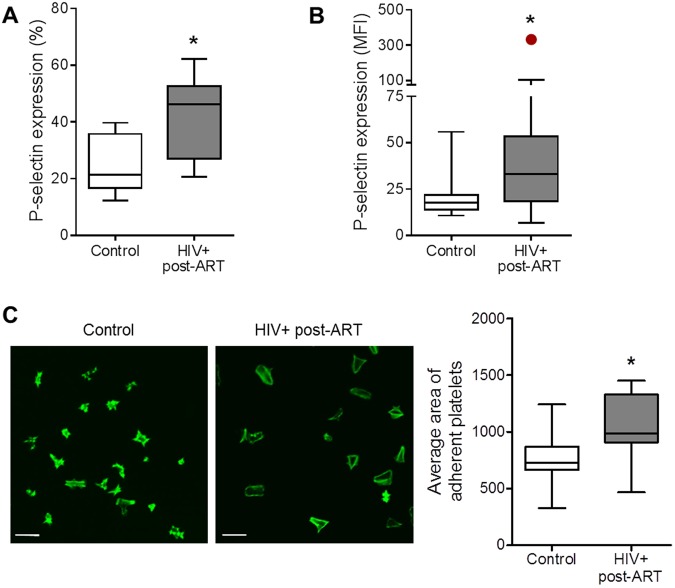
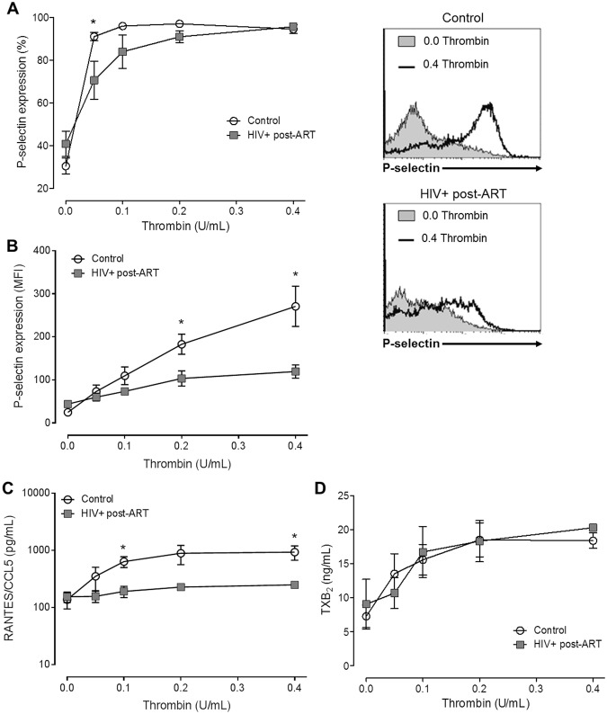
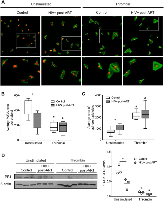
Cardiovascular diseases and thrombotic events became major clinical problems in the combined antiretroviral therapy (cART) era. Although the precise mechanisms behind these clinical problems have not been fully elucidated, a persistent pro-inflammatory state plays a central role. As platelets play important roles on both, thrombus formation and inflammatory/immune response, we aimed at investigating platelet function in HIV-infected subjects virologically controlled through cART. We evaluate parameters of activation, mitochondrial function and activation of apoptosis pathways in platelets from 30 HIV-infected individuals under stable cART and 36 healthy volunteers. Despite viral control achieved through cART, HIV-infected individuals exhibited increased platelet activation as indicated by P-selectin expression and platelet spreading when adhered on fibrinogen-coated surfaces. Platelets from HIV-infected subjects also exhibited mitochondrial dysfunction and activation of apoptosis pathways. Finally, thrombin stimuli induced lower levels of P-selectin translocation and RANTES secretion, but not TXA2 synthesis, in platelets from HIV-infected individuals compared to control; and labeling of platelet alpha granules showed reduced granule content in platelets from HIV-infected individuals when compared to healthy subjects. In summary, platelets derived from HIV-infected individuals under stable cART exhibit a phenotype of increased activation, activation of the intrinsic pathway of apoptosis and undermined granule secretion in response to thrombin.


Conflict of interest statement

The authors declare no competing interests.

Figures

Figure 1
Figure 1
Increased platelet activation in HIV-infected individuals under stable cART. (A,B) The percentage of platelets with P-selectin surface expression (A) and the mean fluorescence intensity (MFI) for P-selectin labeling on platelets (B) were assessed in freshly-isolated platelets from healthy subjects (control) or from HIV-infected individuals under viral control (undetectable HIV-1 viral load in the peripheral blood). (C) Average area of spontaneous spreading on fibrinogen-coated surfaces by platelets from control or HIV-1-infected subjects labeled with phalloidin. Images are representative of eight HIV-infected individuals and healthy volunteers analyzed in parallel. Scale bars represent 10 µm. The horizontal lines on the box plots represent the median, the boxes limits represent the interquartile ranges and the whiskers indicate 5–95 percentile. The dot in panel B represents an outlier (p < 0.05 in Grubbs’ test) that was excluded from statistical analysis. The asterisk (*) signifies p < 0.05 compared to healthy volunteers.
Figure 2
Figure 2
Mitochondrial dysfunction and apoptosis in platelets from HIV-infected individuals under stable cART. (A) The mean fluorescence intensity (MFI) for TMRE indicating the mitochondrial membrane potential (∆ψm), (B) the MFI for MitoSoxRed indicating mitochondrial generation of reactive oxygen species (ROSm), (C) the percentage of platelets that bind annexin V, and (D) the MFI for the probe FLICA indicating caspase-9 activation were assessed in platelets freshly-isolated from healthy volunteers (control) or from HIV-infected individuals under viral control (HIV-1 viral load < 50 copies/ml in the peripheral blood). Box plots: The horizontal lines on the box plots represent the median, boxes limits indicate interquartile ranges and the whiskers indicate 5–95 percentile. The asterisk (*) signify p < 0.05 compared to healthy volunteers.
Figure 3
Figure 3
Platelet response to thrombin in platelets from HIV-infected individuals under stable cART and healthy volunteers. (A,B) The percentage of platelets with P-selectin surface expression (A) and the mean fluorescence intensity (MFI) for P-selectin labeling (B) on platelets from healthy subjects (control) or from HIV-infected individuals under viral control (HIV-1 viral load < 50 copies/ml in the peripheral blood) after stimulation with thrombin (0.05 U/mL, 0.1 U/mL, 0.2 U/mL and 0.4 U/mL). (C,D) The concentration of RANTES (C) or thromboxane B2 (D) in the supernatant of platelets from control or HIV-1-infected subjects stimulated with each concentration of thrombin. The dots indicate the mean ± standard error of the mean of 5–10 HIV-infected individuals or healthy volunteers. Representative histograms are shown. The asterisk (*) signifies p < 0.05 between platelets from control or healthy volunteers stimulated with the same concentration of thrombin.
Figure 4
Figure 4
Thrombin-induced platelet spreading and degranulation in HIV-infected individuals under stable cART and healthy volunteers. (A) Platelets from healthy subjects (control) or HIV-infected individuals under viral control (HIV-1 viral load < 50 copies/ml in the peripheral blood) were stimulated with thrombin (0,0 or 0,4 U/mL), adhered to fibrinogen-coated surfaces and labeled with phalloidin (green) and wheat germ agglutinin (WGA – red) as described in methods. (B) Average area of WGA labeling per platelet number in each field for unstimulated or thrombin-stimulated platelets from control or HIV-1-infected subjects. (C) Average area of spontaneous or thrombin-induced platelet spreading on fibrinogen-coated surfaces in each condition. Images are representative of eight HIV-infected individuals and healthy volunteers analyzed in parallel. Scale bars represent 10 µm. The horizontal lines on the box plots represent the median, boxes limits indicate interquartile ranges and the whiskers indicate 5–95 percentile. (D) Western blot analysis for PF4/CXCL4 and β-actin in platelets from three healthy volunteers and three HIV-infected subjects under stable cART that were kept unstimulated or stimulated with 0.4 U/mL of thrombin. Dots represent the band intensity of PF4 corrected by β-actin expression in each experimental condition. Horizontal lines represent mean. The asterisk (*) signify p < 0.05 compared to healthy volunteers and # means p < 0.05 compared to unstimulated platelets.

References

    1. Lewden C, et al. HIV-infected adults with a CD4 cell count greater than 500 cells/mm3 on long-term combination antiretroviral therapy reach same mortality rates as the general population. J Acquir Immune Defic Syndr. 2007;46(1):72. - PubMed
    1. Dore GJ, et al. Impact of highly active antiretroviral therapy on individual AIDS-defining illness incidence and survival in Australia. J Acquir Immune Defic Syndr. 2002;29(4):388. doi: 10.1097/00126334-200204010-00010. - DOI - PubMed
    1. Marin B, et al. Non-AIDS-defining deaths and immunodeficiency in the era of combination antiretroviral therapy. AIDS. 2009;23(13):1743. doi: 10.1097/QAD.0b013e32832e9b78. - DOI - PMC - PubMed
    1. Hasse B, et al. Morbidity and aging in HIV-infected persons: the Swiss HIV cohort study. Clin Infect Dis. 2011;53(11):1130. doi: 10.1093/cid/cir626. - DOI - PubMed
    1. Casper C. The increasing burden of HIV-associated malignancies in resource-limited regions. Annu Rev Med. 2011;62:157. doi: 10.1146/annurev-med-050409-103711. - DOI - PubMed

Publication types

MeSH terms