Review
. 2018 Dec;16(12):745-759.
doi: 10.1038/s41579-018-0089-x.

The oral microbiota: dynamic communities and host interactions

Affiliations
Review

The oral microbiota: dynamic communities and host interactions

Richard J Lamont et al. Nat Rev Microbiol. 2018 Dec.

Abstract

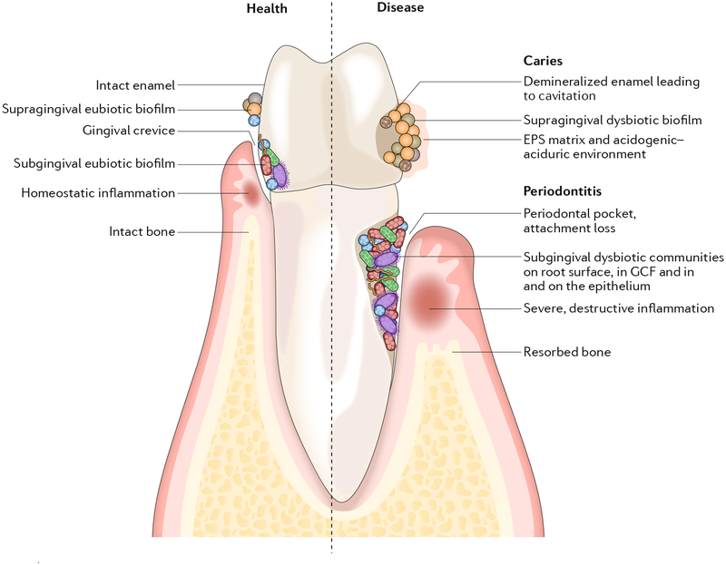
The dynamic and polymicrobial oral microbiome is a direct precursor of diseases such as dental caries and periodontitis, two of the most prevalent microbially induced disorders worldwide. Distinct microenvironments at oral barriers harbour unique microbial communities, which are regulated through sophisticated signalling systems and by host and environmental factors. The collective function of microbial communities is a major driver of homeostasis or dysbiosis and ultimately health or disease. Despite different aetiologies, periodontitis and caries are each driven by a feedforward loop between the microbiota and host factors (inflammation and dietary sugars, respectively) that favours the emergence and persistence of dysbiosis. In this Review, we discuss current knowledge and emerging mechanisms governing oral polymicrobial synergy and dysbiosis that have both enhanced our understanding of pathogenic mechanisms and aided the design of innovative therapeutic approaches for oral diseases.


Conflict of interest statement

Competing interests

The authors declare no competing interests.

Figures

Fig. 1 |
Fig. 1 |. Biogeography of oral microbiota colonization in the diverse habitats of the oral cavity.
Microbial colonization occurs on all available surfaces, and microorganisms can also penetrate epithelial tissues and cells. The microbiota assembles into biofilm communities on the abiotic and biotic surfaces. In health (left), eubiotic biofilms maintain a homeostatic balance with the host. In disease (right), caries and periodontitis ensue when biofilms become dysbiotic, resulting in increased levels and duration of low pH challenge and the induction of destructive inflammatory responses, respectively. EPS, extracellular polymeric substance; GCF, gingival crevicular fluid.
Fig. 2 |
Fig. 2 |. Interactions among bacterial species that affect nososymbiocity.
Oral bacteria interact through multiple pathways that can be separated both spatially and temporally. a | Streptococcus gordonii and Streptococcus parasanguinis produce hydrogen peroxide, to which Aggregatibacter actin-omycetemcomitans responds by activation of the OxyR transcriptional regulator; consequently, transcription of both apiA and katA is elevated. Higher levels of the ApiA surface protein increases complement resistance and will also potentially induce intracellular invasion and pro-inflammatory cytokine production. KatA (a catalase) degrades the hydrogen peroxide produced by both streptococci and neutrophils, thus protecting A. actino-mycetemcomitans from oxidative damage,,–. OxyR also regulates production of dispersin B (DspB), an enzyme that degrades the biofilm matrix and facilitates dispersal of A. actinomycetemcomitans. Hydrogen peroxide increases the bioavailability of oxygen, and, in response, A. actinomycetemcomitans shifts from a primarily fermentative to a respiratory metabolism, an interaction referred to as cross respiration. Respiratory metabolism enhances the growth and fitness of A. actinomycetemcomitans in vivo. Transport of streptococcal lactate into A. actinomycetemcomitans through the proton-driven lactate permease (LctP) leads to conversion to pyruvate by lactate dehydrogenase (LctD). Pyruvate suppresses autophosphorylation of E1, which then decreases uptake of phosphotransferase system (PTS) carbohydrates such as glucose. Preferential utilization of lactate through this carbon resource partitioning gives a competitive advantage to A. actinomycetemcomitans in the presence of organisms that can metabolize glucose more efficiently. Communities of A. actinomycetemcomitans and S. gordonii also become restricted for iron. The Fur transcriptional regulator of A. actinomycetemcomitans responds to iron limitation and induces upregulation of the gene encoding DspB, which will release A. actinomycetemcomitans from biofilms. A. actinomycetemcomitans responses to both oxidative stress and iron restriction thus involve DspB activity and re-localization, and in vivo A. actinomycetemcomitans maintains an optimal distance from streptococci in communities that are synergistically virulent. b | Interactions between Porphyromonas gingivalis and S. gordonii resulting from metabolite (4-amino benzoate (pABA)) perception (left) and direct contact (right). pABA secreted by S. gordonii inactivates the P. gingivalis tyrosine phosphatase Ltp1. Dephosphorylation and inactivation of the tyrosine kinase Ptk1 is thus reduced. Ptk1 phosphorylates and inactivates the transcription factor CdhR, which is a repressor of the mfa1 gene. Ptk1 activity also converges on expression of the fimA gene. Expression of both fimbrial adhesins is increased, and in this mode P. gingivalis is primed for attachment to S. gordonii. However, nososymbiocity is reduced, and pABA-treated P. gingivalis are less pathogenic in animal models. Engagement of Mfa1 with the streptococcal SspA or SspB surface protein increases Ltp1 and reverses information flow through the Ltp1-Ptk1 axis. In addition, Mfa1-SspA/SspB binding suppresses expression of chorismate binding enzyme (Cbe), which is responsible for pABA production. Prolonged physical interaction between P. gingivalis and S. gordonii leads to increased nososymbiocity, and dual infection of animal models causes more alveolar bone loss than P. gingivalis infection alone–,,. c | Streptococcus cristatus arginine deiminase (ArcA) interacts with the P. gingivalis surface protein RagB. Signal transduction results in downregulation of genes encoding the FimA and Mfa1 component fimbriae along with the arginine-specific (RgpA or RgpB) and lysine-specific (Kgp) gingipain proteinases. Adhesion, biofilm formation, epithelial cell invasion and degradation of cytokines are consequently reduced and nososymbiocity is suppressed. Part a adapted with permission from REF, Wiley-VCH.
Fig. 3 |
Fig. 3 |. Diet-microbiota interactions trigger the assembly of cariogenic biofilm microenvironment.
In the oral microbial community on tooth surfaces, social interactions begin with primary colonizers that can rapidly attach and then co-adhere with later colonizers. Microorganisms can interact physically and metabolically to determine the initial biofilm community. Both antagonistic and cooperative interactions can occur, and these dynamically change according to the host diet, and other factors such as salivary dysfunction, fluoride exposure and oral hygiene. In particular, dietary sucrose provides a substrate for extracellular polysaccharide production and synthesis of organic acids by acidogenic microorganisms. The extracellular matrix, which also contains other biomolecules (extracellular DNA (eDNA) and bacterial or host-derived proteins), provides a multi-functional scaffold for spatial organization, mechanical coherence and interbacterial interactions. The matrix can trap or sequester substances, which, in combination with diffusion-modifying properties, can generate a variety of chemical and protective microenvironments. Biofilms thus become persistently adhered to the surface and recalcitrant to antimicrobial action. S. mutans has a key pathogenic role as an EPS-matrix producer, acidogenic and aciduric organism. With frequent dietary sugar exposure, continued bacterial metabolism of carbohydrates and reduced accessibility to salivary buffering systems causes the microenvironment within the matrix to become increasingly and constantly acidic. As the biofilm accumulates, the microenvironment also becomes progressively anaerobic (hypoxic). In a feedforward loop, microbial diversity decreases as an aciduric microbiota predominates. If the biofilm is not removed, persistent low-pH conditions at the tooth-biofilm interface shift the demineralization-remineralization balance towards net mineral loss from the tooth enamel, leading to the development of a carious lesion. EPS, extracellular polymeric substance. Adapted with permission from REF, Cell Press.
Fig. 4 |
Fig. 4 |. Reciprocally reinforced interactions between dysbiosis and inflammation drive chronic periodontitis.
Colonization by keystone pathogens (for example, Porphyromonas gingivalis) aided by accessory pathogens (for example, Streptococcus gordonii) leads to impaired innate host defence and promotion of inflammation (for example, by subverting complement-Toll-like receptor (TLR) crosstalk in neutrophils and other myeloid cells),,,. These alterations contribute to the emergence of dysbiosis (quantitative and compositional changes in the periodontal microbiota). Inflammation worsens dysbiosis by increasing the flow of gingival crevicular fluid (GCF), which, as a result of inflammatory tissue destruction, carries degraded collagen and haem-containing compounds into the gingival crevice, where dysbiotic communities develop. These molecules are selectively used by proteolytic and asaccharolytic bacteria with iron-acquisition capacity. By contrast, health-associated (eubiotic) species cannot capitalize on the new environmental conditions and are outcompeted. This imbalance drives dysbiosis, which further exacerbates inflammation, culminating in periodontitis in susceptible individuals. The ability of inflammation and dysbiosis to positively reinforce each other in a self-sustained feedforward loop may contribute to the chronicity of periodontitis.
Fig. 5 |
Fig. 5 |. P. gingivalis induces dysbiosis by impairing innate host defences while promoting inflammatory responses in phagocytic cells.
Porphyromonas gingivalis expresses cell-surface molecules that activate the Toll-like receptor 2 (TLR2)-TLR1 complex and secretes enzymes (HRgpA and RgpB gingipains) that act on the complement component C5 to generate high local concentrations of C5a, a ligand of complement C5a receptor 1 (C5aR1). The bacterium can thus co-activate C5aR1 and TLR2 in phagocytic cells such as neutrophils and macrophages. In both of these myeloid cell types, P gingivalis can bypass MyD88 and thus prevent the associated bactericidal activity,, which in neutrophils is possibly mediated by downstream activation of IRAK4-dependent granule exocytosis. In neutrophils, the inactivation of MyD88 involves its ubiquitylation via the E3 ubiquitin ligase SMURF1 and subsequent proteasomal degradation. Although MyD88-dependent inflammation is blocked by P. gingivalis, this organism induces PI3K-dependent inflammatory cytokines in both neutrophils and macrophages,. Similarly, in both cell types, P. gingivalis-induced activation of PI3K leads to inhibition of phagocytosis,. In neutrophils, this activity is mediated by the ability of PI3K to suppress RhoA GTPase and actin polymerization. Intriguingly, even within those macrophages that do manage to phagocytose P. gingivalis bacteria, PI3K signalling suppresses phago-lysosomal maturation, thereby preventing pathogen destruction. These tactics compromise innate immunity and promote inflammation that leads to the selective expansion of inflammophilic pathobionts. Conversely, inhibition of C5aR1, TLR2 or PI3K reverses dysbiotic inflammation and periodontitis in mice,. MAL, MyD88-adaptor-like.
Fig. 6 |
Fig. 6 |. Localized chemokine paralysis.
Oral pathobionts such as Fusobacterium nucleatum are recognized by Toll-like receptors (TLRs) on epithelial cell surfaces, which leads to the activation of pro-inflammatory signalling pathways. The keystone pathogen Porphyromonas gingivalis can manipulate these pathways and cause a targeted and precise reduction in the production of specific chemokines. Inactivation of STAT1 by P gingivalis leads to reduced expression of CXCL10, which is controlled by the IRF1 transcription factor. Intracellularly, P. gingivalis secretes SerB, a serine phosphatase that specifically dephosphorylates the serine 536 residue of the p65 NF-κB subunit, thus inhibiting formation and nuclear translocation of NF-κB-p65 homodimers. Transcription of the IL8 gene is reduced and the IL-8 neutrophil gradient is disrupted. These chemokine paralysis activities will be localized to tissue adjacent to, or containing, P. gingivalis, and in animal models supersede the effects of community pathobionts. The continuous recalibration of host cell signalling pathways also limits the temporal extent of the phenomenon, which may contribute to the cyclical nature of periodontal tissue destruction. Adapted with permission from REF, Cell Press.

Similar articles

Cited by

References

    1. Abusleme L et al. The subgingival microbiome in health and periodontitis and its relationship with community biomass and inflammation. ISME J. 7, 1016–1025 (2013).

      This study documents alterations in subgingival microbial communities that underpin the development of periodontitis and describes the relationship between clinical inflammation and the disease-associated microbiome.

    1. Griffen AL et al. Distinct and complex bacterial profiles in human periodontitis and health revealed by 16S pyrosequencing. ISME J. 6, 1176–1185 (2012).

      This landmark study establishes the complexity of the periodontal microbial community and the demarcation between health and disease.

    1. Rosan B & Lamont RJ Dental plaque formation. Microbes Infect. 2, 1599–1607 (2000). - PubMed
    1. Aas JA, Paster BJ, Stokes LN, Olsen I & Dewhirst FE Defining the normal bacterial flora of the oral cavity. J. Clin. Microbiol 43, 5721–5732 (2005). - PMC - PubMed
    1. Dewhirst FE et al. The human oral microbiome. J. Bacteriol 192, 5002–5017 (2010).

      References 4 and 5 are the basis of our current understanding of the diversity of the oral microbiome.

Publication types