. 2018 Oct 9;8(1):14990.
doi: 10.1038/s41598-018-33331-z.

Peptide-oligonucleotide conjugates exhibiting pyrimidine-X cleavage specificity efficiently silence miRNA target acting synergistically with RNase H

Affiliations

Peptide-oligonucleotide conjugates exhibiting pyrimidine-X cleavage specificity efficiently silence miRNA target acting synergistically with RNase H

O A Patutina et al. Sci Rep. .

Abstract

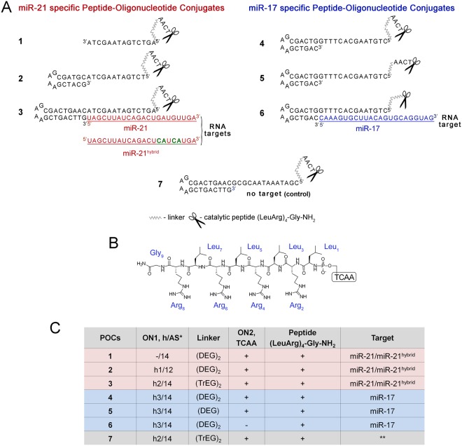
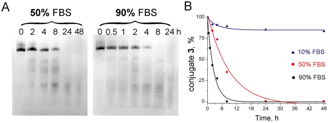
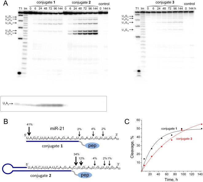
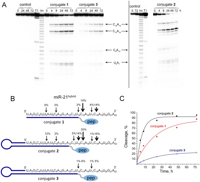
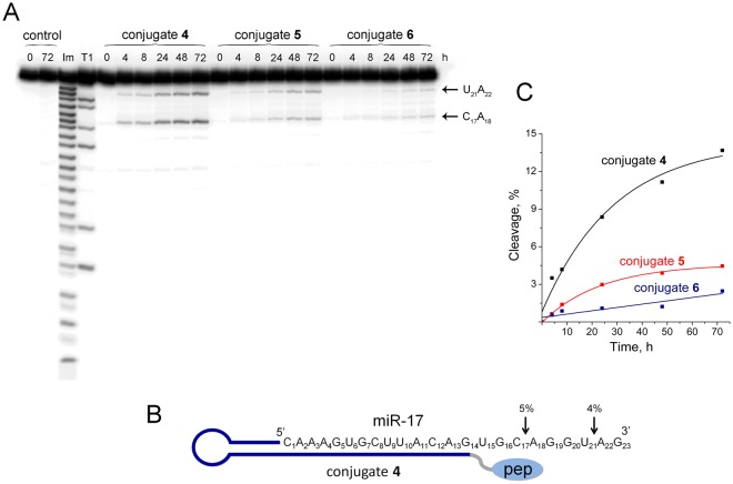
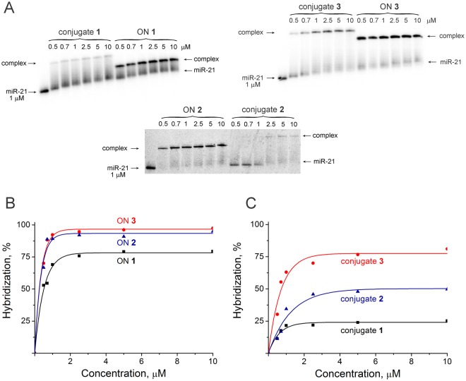
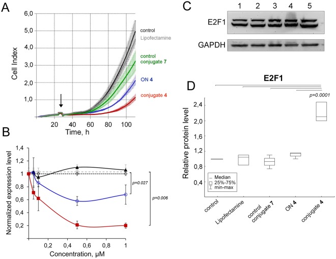
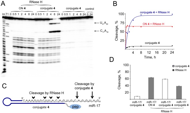
Taking into account the important role of miRNA in carcinogenesis, oncogenic miRNAs are attractive molecules for gene-targeted therapy. Here, we developed a novel series of peptide-oligonucleotide conjugates exhibiting ribonuclease activity targeted to highly oncogenic miRNAs miR-21 and miR-17. When designing the conjugates, we enhanced both nuclease resistance of the targeted oligodeoxyribonucleotide by introducing at its 3'-end mini-hairpin structure displaying high thermostability and robustness against nuclease digestion and the efficiency of its functioning by attachment of the catalytic construction (amide)NH2-Gly(ArgLeu)4-TCAA displaying ribonuclease activity to its 5'-end. Designed miRNases efficiently cleaved miRNA targets, exhibiting Pyr-X specificity, and cleavage specificity had strong dependence on the miRNA sequence in the site of peptide location. In vitro, designed miRNases do not prevent cleavage of miRNA bound with the conjugate by RNase H, and more than an 11-fold enhancement of miRNA cleavage by the conjugate is observed in the presence of RNase H. In murine melanoma cells, miRNase silences mmu-miR-17 with very high efficiency as a result of miR-17 cleavage by miRNase and by recruited RNase H. Thus, miRNases provide a system of double attack of the miRNA molecules, significantly increasing the efficiency of miRNA downregulation in the cells in comparison with antisense oligonucleotide.


Conflict of interest statement

The authors declare no competing interests.

Figures

Figure 1
Figure 1
The structure of anti-miRNA peptide-oligonucleotide conjugates (POCs). (A) Schematic representation of a general complex of RNA target and peptide-oligonucleotide conjugate. (B) The chemical structure of the peptide (LeuArg)4-Gly-NH2. The peptide was conjugated via the N-terminus to the short oligonucleotide TCAA, which was attached via diethylene glycol (DEG) or tetraethylene glycol (TrEG) linker to the 5′-terminal phosphate of the antisense oligonucleotide. (C) Structural components of conjugates. Designations: ON1, h/AS – oligonucleotide in the conjugate containing hairpin (h) and sequence complementary to miRNA (AS); h1 and h3 – hairpins with 6 b.p. stem, h2 - hairpin with 9 b.p. stem; DEG – diethylene glycol; TrEG - tetraethylene glycol; ON2 – TCAA; * - conjugate 7 (control conjugate) was targeted to luciferase mRNA.
Figure 2
Figure 2
Stability of conjugate 3 in DMEM supplemented with 10–90% fetal bovine serum. (A) Stability of the conjugate 3 in DMEM supplemented with 50% (left panel) and with 90% FBS (right panel). Autoradiographs of 12% polyacrylamide/8 M urea gel. (B) The time course of conjugate 3 degradation in 10 (triangles), 50 (circles) and 90% (squares) FBS.
Figure 3
Figure 3
Cleavage of 5′-[32P]-miR-21 by conjugates 1, 2, and 3. (A) Autoradiograph of 18% polyacrylamide/8 M urea gel, showing the pattern of miR-21 cleavage by the conjugates. Lanes Im and T1 — imidazole ladder and partial RNA digestion with RNase T1, respectively; control — RNA incubated in the absence of conjugates for 0 and 144 h. miR-21 (1 µM) and conjugates (100 µM) were incubated at 37 °C for 0–144 h. The conjugate type and incubation time are shown at the top. The image enclosed by lines is part of the upper left image but scanned as separate file. (B) Positions of miR-21 cleavage by conjugates 1 and 2. The percentage of cleavage at specific sites in 144 h is indicated above the arrows; pep — catalytic construction. (C). Kinetics of miR-21 cleavage by conjugates 1 and 2.
Figure 4
Figure 4
Cleavage of 5′-[32P]-miR-21hybrid by conjugates 1, 2, and 3. (A) Autoradiograph of 18% polyacrylamide/8 M urea gel showing the pattern of miR-21hybrid cleavage by the conjugates. Lanes Im and T1 — imidazole ladder and partial RNA digestion with RNase T1, respectively; control — RNA incubated in the absence of conjugates for 0–72 h. miR-21 (1 µM) and conjugates (20 µM) were incubated at 37 °C for 0–72 h. The conjugate type and incubation time are shown at the top. The images enclosed by lines are parts of the same gel. (B) Positions of miR-21hybrid cleavage by conjugates. The percentage of cleavage at specific sites in 24 h is indicated above the arrows; pep — catalytic construction. (C). Kinetics of miR-21hybrid cleavage by conjugates.
Figure 5
Figure 5
Cleavage of 5′-[32P]-miR-17 by conjugates 4, 5, and 6. (A) Autoradiograph of 18% polyacrylamide/8 M urea gel showing the pattern of miR-17 cleavage by the conjugates. Lanes Im and T1 — imidazole ladder and partial RNA digestion with RNase T1, respectively; control — RNA incubated in the absence of conjugates for 0 and 72 h. miR-17 (1 µM) and conjugates (20 µM) were incubated at 37 °C for 0–72 h. The conjugate type and incubation time are shown at the top. The images enclosed by lines are parts of the same gel. (B) Positions of miR-17 cleavage by conjugate 4. The percentage of cleavage at specific sites in 24 h is indicated above by arrows; pep — catalytic construction. (C). Kinetics of miR-17 cleavage by conjugates.
Figure 6
Figure 6
Hybridization efficiency of conjugates 13 and corresponding oligonucleotides 13 (ON 13) with 5′-[32P]-miR-21. (A) Autoradiograph of 15% native PAGE. (B,C). Concentration dependencies of binding of oligonucleotides and conjugates with miR-21.
Figure 7
Figure 7
Biological effect of conjugate 4 in melanoma B16 cells. (A) Real-time analysis of the effect of anti-miR-17 conjugate 4 on the growth rate of B16 melanoma cells. B16 melanoma cells were transfected with conjugate 4, control conjugate 7, oligonucleotide 4 (ON 4) precomplexed with Lipofectamine 2000 at a concentrations of 1 µM. B16 cells without any treatment and B16 treated with Lipofectamine were used as controls. Transfection time is indicated by arrow. The data were statistically processed using the Student’s t-test (two-tailed, unpaired). The results are shown as mean cell index ± standard error. (B) Expression level of miR-17 in tumor cells after transfection with anti-miR-17 conjugate 4. Control conjugate 7, conjugate 4 and oligonucleotide 4 (ON 4) were transfected in complex with Lipofectamine 2000 into melanoma cells B16 in concentration range 0.05–1 µM. In 24 h after transfection RNA was isolated and stem-loop PCR was performed. The expression of miR-17 was normalized to U6. Data were statistically analyzed using one-way ANOVA with post hoc Tukey test. Data are given as mean calculated from three independent experiments ± SEM. Open diamond symbol — control, cells without any treatment; cross — cells treated with Lipofectamine; close triangle — cells treated with control conjugate 7; open circle — cells treated with oligonucleotide 4; close square — cells treated with conjugate 4. (C) Western blot analysis of E2F1 protein level 72 h after transfection. GAPDH served as an internal control. 1 intact melanoma B16 cells; 2 melanoma B16 cells incubated with Lipofectamine 2000; 3, 4, 5 melanoma B16 cells incubated with 1 µM control conjugate 7, antisense oligonucleotide ON 4 and anti-miR-17 conjugate 4, respectively. (D) The bar graph shows the semi-quantitative analysis of the Western blot results for E2F1. Data were statistically analyzed using one-way ANOVA with post hoc Tukey test.
Figure 8
Figure 8
Cleavage of miR-17 by miR-17-specific conjugate 4 and RNase H. (A) Patterns of 5′-[32P]-miR-17 cleavage in complex with conjugate 4 or in complex with oligonucleotide 4 (ON 4) by RNase H. Autoradiograph of 18% polyacrylamide/8 M urea gel. Duplexes of 5′-[32P]-miR-17 (10 μM) and oligonucleotide or conjugate (5 μM) were incubated at 37 °C for 24 h with RNase H (100 U/ml). Lanes Im and T1 — imidazole ladder and partial RNA digestion with RNase T1, respectively; control — RNA was incubated in the absence of oligonucleotide/conjugate and in the presence of RNase H. (B) Kinetics of miR-17 cleavage by conjugates 4, miR-17/ON 4 cleavage by RNase H, and miR-17 cleavage by conjugate 4 together with RNase H. (C) Positions of miR-17 cleavage by RNase H and conjugate 4. Cleavage at specific sites is indicated by arrows; pep — catalytic construction. (D) Diagram showing contribution of conjugate 4 and RNase H in the total cleavage of miR-17 for 8 h. Dark grey bars and white bars show contribution of RNase H and conjugate 4 to the miR-17 cleavage, respectively.

Similar articles

Cited by

References

    1. Hanahan D, Weinberg RA. Hallmarks of cancer: the next generation. Cell. 2011;144:646–674. doi: 10.1016/j.cell.2011.02.013. - DOI - PubMed
    1. Grijalvo S, Alagia A, Jorge AF, Eritja R. Covalent strategies for targeting messenger and non-coding RNAs: an updated review on siRNA, miRNA and antimiR conjugates. Genes (Basel) 2018;9:74. doi: 10.3390/genes9020074. - DOI - PMC - PubMed
    1. Ambros V. The functions of animal microRNAs. Nature. 2004;431:350–355. doi: 10.1038/nature02871. - DOI - PubMed
    1. Bartel DP. MicroRNAs: genomics, biogenesis, mechanism, and function. Cell. 2004;116:281–297. doi: 10.1016/S0092-8674(04)00045-5. - DOI - PubMed
    1. Pillai RS, et al. Inhibition of translational initiation by Let-7 microRNA in human cells. Science. 2005;309:1573–1576. doi: 10.1126/science.1115079. - DOI - PubMed

Publication types

MeSH terms