. 2018 Sep 27:4:30.
doi: 10.1038/s41531-018-0066-0. eCollection 2018.

Ingestion of subthreshold doses of environmental toxins induces ascending Parkinsonism in the rat

Affiliations

Ingestion of subthreshold doses of environmental toxins induces ascending Parkinsonism in the rat

L Anselmi et al. NPJ Parkinsons Dis. .

Abstract

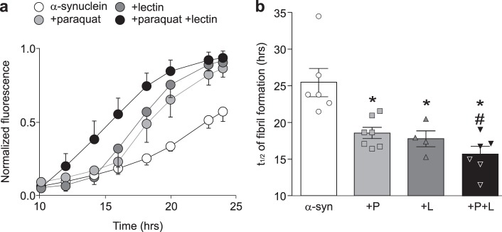
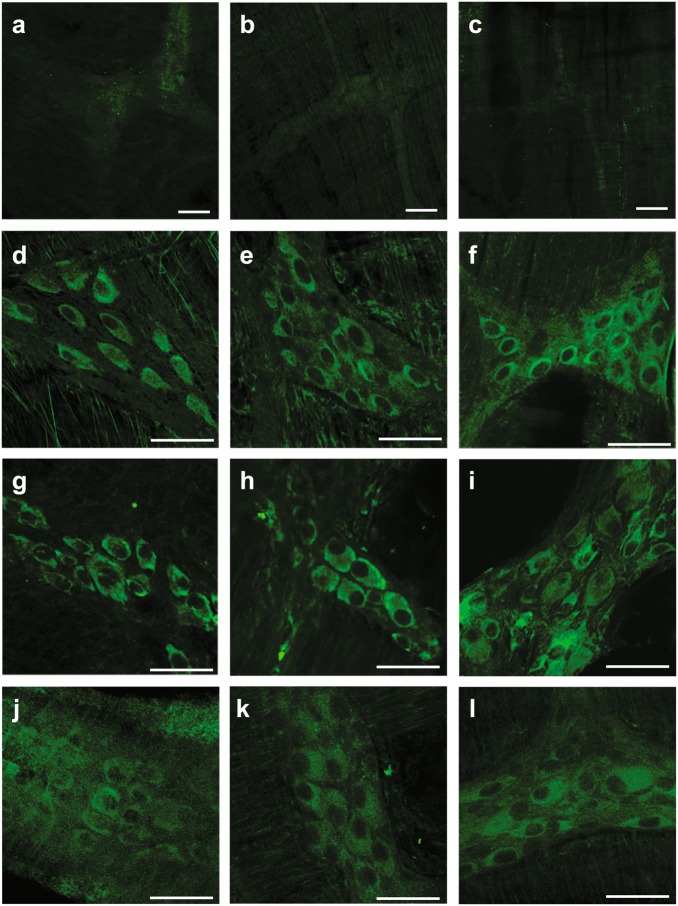
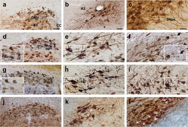
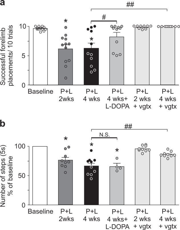
Increasing evidence suggests that environmental neurotoxicants or misfolded α-synuclein generated by such neurotoxicants are transported from the gastrointestinal tract to the central nervous system via the vagus nerve, triggering degeneration of dopaminergic neurons in the substantia nigra pars compacta (SNpc) and causing Parkinson's disease (PD). We tested the hypothesis that gastric co-administration of subthreshold doses of lectins and paraquat can recreate the pathology and behavioral manifestations of PD in rats. A solution containing paraquat + lectin was administered daily for 7 days via gastric gavage, followed by testing for Parkinsonian behavior and gastric dysmotility. At the end of the experiment, brainstem and midbrain tissues were analyzed for the presence of misfolded α-synuclein and neuronal loss in the SNpc and in the dorsal motor nucleus of the vagus (DMV). Misfolded α-synuclein was found in DMV and SNpc neurons. A significant decrease in tyrosine hydroxylase positive dopaminergic neurons was noted in the SNpc, conversely there was no apparent loss of cholinergic neurons of the DMV. Nigrovagally-evoked gastric motility was impaired in treated rats prior to the onset of parkinsonism, the motor deficits of which were improved by l-dopa treatment. Vagotomy prevented the development of parkinsonian symptoms and constrained the appearance of misfolded α-synuclein to myenteric neurons. These data demonstrate that co-administration of subthreshold doses of paraquat and lectin induces progressive, l-dopa-responsive parkinsonism that is preceded by gastric dysmotility. This novel preclinical model of environmentally triggered PD provides functional support for Braak's staging hypothesis of idiopathic PD.


Conflict of interest statement

The authors declare no competing interests.

Figures

Fig. 1
Fig. 1
Incubation with paraquat + lectin increases the rate of α-synuclein fibrillation. a Time course of α-synuclein fibrillation in the presence of α-synuclein alone (white, α-syn, n = 6), paraquat (light gray, P, n = 7), lectin (dark gray, L, n = 4) or a combination of paraquat + lectin (black, P + L, n = 6). b Graphic summary of fibrillation t1/2 for α-synuclein. *p < 0.05 vs. α-synuclein alone; #p < 0.05 vs. paraquat or lectin
Fig. 2
Fig. 2
Paraquat + lectin treatment promotes α-synuclein misfolding in myenteric neurons of the GI tract. Representative micrographs showing 129Ser α-synuclein in the myenteric plexus isolated from the stomach a, d, g, j, the small intestine b, e, h, k, and the large intestine c, f, i, l from control animals (top row), animals sacrificed two (second row) or four (third row) weeks after the end of the gavage with paraquat + lectin, or animals that received subdiaphragmatic vagotomy prior to the paraquat + lectin treatment (bottom row). Calibration bars: 50 μm
Fig. 3
Fig. 3
Misfolded α-synuclein is present in the DMV, the A2 area, and the SNpc after treatment with paraquat and lectin. Representative micrographs showing the co-localization of ChAT- or TH-immunoreactivity and 129Ser α-synuclein-immunoreactivity. Representative micrographs showing the same rostro-caudal level of the DMV in control a, 2 d, and 4 g weeks after the last gavage with paraquat and lectin, or in animals that received subdiaphragmatic vagotomy prior to the treatment j. ChAT-immunoreactivity (brown, a, d, and j; blue in g) and 129Ser α -synuclein (blue in a, d, and j; brown in g); calibration bar: 75 µm. Representative micrographs showing the A2 area in control b, 2 e, and 4 h weeks after the last gavage with paraquat + lectin, or in animals that received subdiaphragmatic vagotomy prior to the treatment k. TH-IR (brown) and 129Ser α -synuclein (blue); calibration bar: 75 µm. Representative micrographs showing the same area of the SNpc in control c, 2 f, and 4 i weeks after the last gavage with paraquat + lectin, or in animals that received subdiaphragmatic vagotomy prior to the treatment l. TH-IR (brown) and 129Ser α -synuclein (blue); calibration bar: 50 µm. Insets are higher magnifications of their respective panels
Fig. 4
Fig. 4
Paraquat and lectin treatment impairs motor activity. Graphic summary showing the motor performance of rats examined with the vibrissae a and stepping b tests following treatment with paraquat + lectin. Note that the motor activity was significantly reduced two weeks after the end of treatment and persisted thereafter. l-dopa pretreatment induced a significant improvement of motor performance assessed with the vibrissae test (n = 12). Animals that received subdiaphragmatic vagotomy (n = 9) prior to the treatment did not show any motor impairment. *p < 0.05 vs. baseline
Fig. 5
Fig. 5
Treatment with paraquat + lectin induces loss of TH-positive neurons in the SNpc. Representative images of TH-positive neurons in SNpc of control a and treated animals b. Insets are higher magnifications of the boxed areas in the respective panels. Calibration bar: 100 µm. Graphic summary of TH-IR neuronal number in both the left c and right d SNpc of control (white) and P + L treated (black) rats. Note that a significant loss of neurons was detected in SNpc of animals 4 weeks after the last gavage of paraquat + lectin (n = 3 for controls and 4 for P + L 4 weeks, respectively; *p < 0.05 vs. control). d Graphic summary showing that the mean estimate number of TH-IR neurons is not significantly different from the mean estimate number of CV-positive neurons in the SNpc of P + L treated animals (n = 4)
Fig. 6
Fig. 6
Treatment with paraquat and lectin induces impairment of the nigro–vagal pathway. a Representative recordings from the gastric antrum showing that the increase of tone and motility following microinjection of NMDA in the left SNpc was reduced progressively at 2 and 4 weeks after the last gavage treatment. b Bottom panel: graphic summaries showing that 2 or 4 weeks after the paraquat and lectin treatment, microinjection of NMDA in the left SNpc increased gastric antrum tone (n = 9, 6, 10 for control, P + L 2 weeks and P + L 4 weeks, respectively) and motility (n = 7, 7, 9 for control, P + L 2 weeks and P + L 4 weeks, respectively) to a significantly lesser extent than in controls (*p < 0.05 vs. control). c Representative recordings from the gastric antrum showing that the increase of tone and motility following microinjection of TRH (1 pmole/60 nl) in the DMV was reduced progressively at 2 and 4 weeks after the last gavage treatment. d Graphic summaries showing that, 2 or 4 weeks after the paraquat and lectin treatment, microinjection of TRH (0.1–3 pmoles/60 nl) in the DMV increased gastric antrum tone and motility to a significantly lesser extent than in controls (*p < 0.05 vs. control)

References

    1. Goedert M, Spillantini MG, Del TK, Braak H. 100 years of Lewy pathology. Nat. Rev. Neurol. 2013;9:13–24. doi: 10.1038/nrneurol.2012.242. - DOI - PubMed
    1. Travagli RA, Anselmi L. Vagal neurocircuitry and its influence on gastric motility. Nat. Rev. Gastroenterol. Hepatol. 2016;13:389–401. doi: 10.1038/nrgastro.2016.76. - DOI - PMC - PubMed
    1. Pellegrini C, et al. Gastric motor dysfunctions in Parkinson’s disease: current pre-clinical evidence. Parkinsonism Relat. Disord. 2015;21:1407–1414. doi: 10.1016/j.parkreldis.2015.10.011. - DOI - PubMed
    1. Fasano A, Visanji NP, Liu LW, Lang AE, Pfeiffer RF. Gastrointestinal dysfunction in Parkinson’s disease. Lancet Neurol. 2015;14:625–639. doi: 10.1016/S1474-4422(15)00007-1. - DOI - PubMed
    1. Rao M, Gershon MD. The bowel and beyond: the enteric nervous system in neurological disorders. Nat. Rev. Gastroenterol. Hepatol. 2016;13:517–528. doi: 10.1038/nrgastro.2016.107. - DOI - PMC - PubMed