

Target identification for the diagnosis and intervention of vulnerable atherosclerotic plaques beyond 18F-fluorodeoxyglucose positron emission tomography imaging: promising tracers on the horizon
- PMID: 30302506
- PMCID: PMC6267660
- DOI: 10.1007/s00259-018-4176-z
Target identification for the diagnosis and intervention of vulnerable atherosclerotic plaques beyond 18F-fluorodeoxyglucose positron emission tomography imaging: promising tracers on the horizon
Abstract
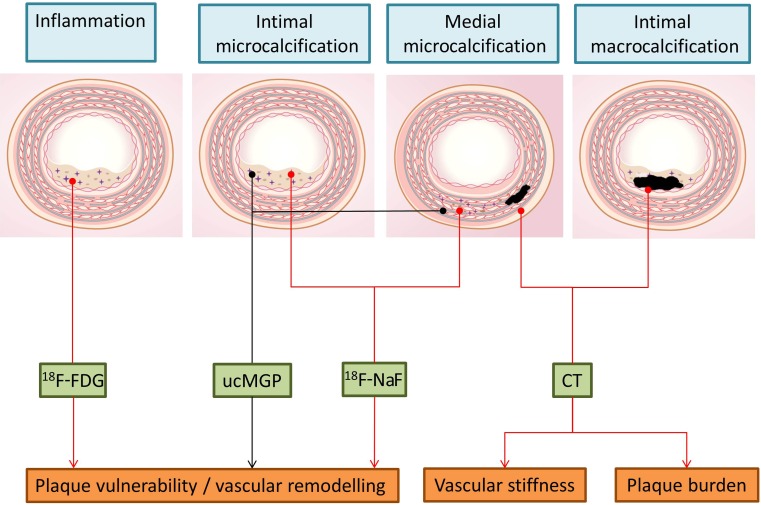
Cardiovascular disease is the major cause of morbidity and mortality in developed countries and atherosclerosis is the major cause of cardiovascular disease. Atherosclerotic lesions obstruct blood flow in the arterial vessel wall and can rupture leading to the formation of occlusive thrombi. Conventional diagnostic tools are still of limited value for identifying the vulnerable arterial plaque and for predicting its risk of rupture and of releasing thromboembolic material. Knowledge of the molecular and biological processes implicated in the process of atherosclerosis will advance the development of imaging probes to differentiate the vulnerable plaque. The development of imaging probes with high sensitivity and specificity in identifying high-risk atherosclerotic vessel wall changes and plaques is crucial for improving knowledge-based decisions and tailored individual interventions. Arterial PET imaging with 18F-FDG has shown promising results in identifying inflammatory vessel wall changes in numerous studies and clinical trials. However, due to its limited specificity in general and its intense physiological uptake in the left ventricular myocardium that impair imaging of the coronary arteries, different PET tracers for the molecular imaging of atherosclerosis have been evaluated. This review describes biological, chemical and medical expertise supporting a translational approach that will enable the development of new or the evaluation of existing PET tracers for the identification of vulnerable atherosclerotic plaques for better risk prediction and benefit to patients.
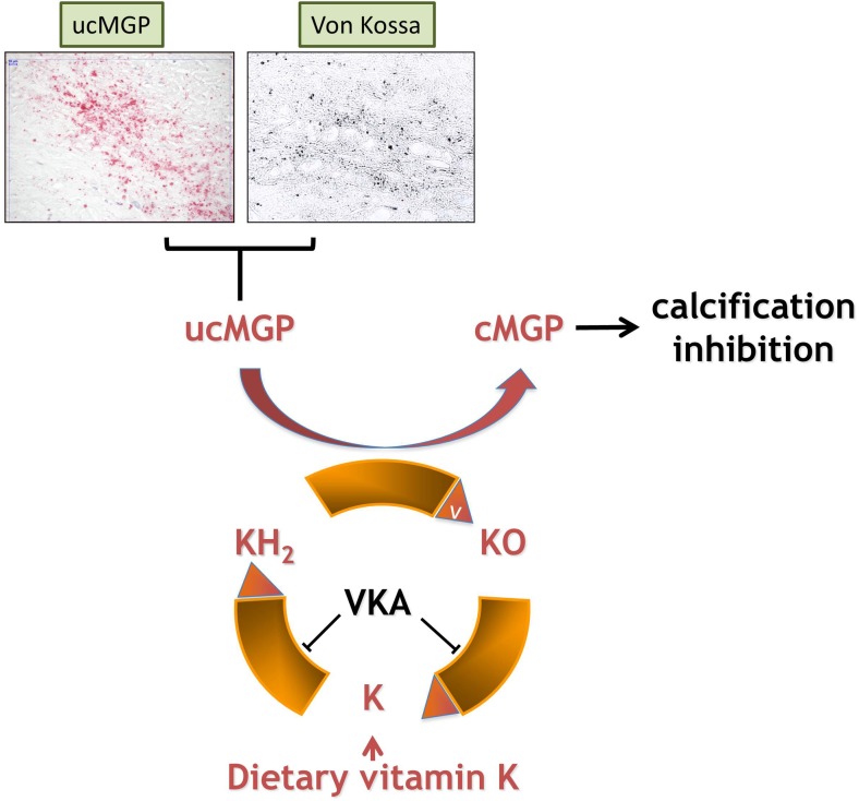
Keywords: Atherosclerotic plaque; Cardiovascular disease; PET tracers; Vascular calcification; Vascular smooth muscle cells.
Conflict of interest statement
Conflict of interest
None.
Studies with human participants or animals
This article does not describe any studies with human participants or animals performed by any of the authors.
Figures
Similar articles
-
Vascular Inflammation in Subclinical Atherosclerosis Detected by Hybrid PET/MRI.J Am Coll Cardiol. 2019 Apr 2;73(12):1371-1382. doi: 10.1016/j.jacc.2018.12.075. J Am Coll Cardiol. 2019. PMID: 30922468
-
Targeted PET/CT imaging of vulnerable atherosclerotic plaques: microcalcification with sodium fluoride and inflammation with fluorodeoxyglucose.Curr Cardiol Rep. 2013 Jun;15(6):364. doi: 10.1007/s11886-013-0364-4. Curr Cardiol Rep. 2013. PMID: 23605466 Review.
-
High-risk plaque features can be detected in non-stenotic carotid plaques of patients with ischaemic stroke classified as cryptogenic using combined (18)F-FDG PET/MR imaging.Eur J Nucl Med Mol Imaging. 2016 Feb;43(2):270-279. doi: 10.1007/s00259-015-3201-8. Epub 2015 Oct 3. Eur J Nucl Med Mol Imaging. 2016. PMID: 26433367
-
Potential of α7 nicotinic acetylcholine receptor PET imaging in atherosclerosis.Methods. 2017 Nov 1;130:90-104. doi: 10.1016/j.ymeth.2017.06.008. Epub 2017 Jun 8. Methods. 2017. PMID: 28602809 Review.
-
Imaging of atherosclerotic aorta of rabbit model by detection of plaque inflammation with fluorine-18 fluorodeoxyglucose positron emission tomography/computed tomography.Chin Med J (Engl). 2011 Mar;124(6):911-7. Chin Med J (Engl). 2011. PMID: 21518602
Cited by
-
Radionuclide imaging of inflammation in atherosclerotic vascular disease among people living with HIV infection: current practice and future perspective.Eur J Hybrid Imaging. 2019 Apr 11;3(1):5. doi: 10.1186/s41824-019-0053-7. Eur J Hybrid Imaging. 2019. PMID: 34191183 Free PMC article. Review.
-
The Role of Vascular Smooth Muscle Cells in Arterial Remodeling: Focus on Calcification-Related Processes.Int J Mol Sci. 2019 Nov 14;20(22):5694. doi: 10.3390/ijms20225694. Int J Mol Sci. 2019. PMID: 31739395 Free PMC article. Review.
-
Targeting vulnerable atherosclerotic plaque via PET-tracers aiming at cell-surface overexpression of somatostatin receptors.Biomed Rep. 2020 Sep;13(3):9. doi: 10.3892/br.2020.1316. Epub 2020 Jun 16. Biomed Rep. 2020. PMID: 32765848 Free PMC article.
-
18F-fluorodeoxyglucose positron emission tomography for the detection of inflammatory lesions of the arterial vessel walls in Wistar rats.Exp Ther Med. 2021 Apr;21(4):370. doi: 10.3892/etm.2021.9801. Epub 2021 Feb 19. Exp Ther Med. 2021. PMID: 33732343 Free PMC article.
-
Molecular Imaging and Non-molecular Imaging of Atherosclerotic Plaque Thrombosis.Front Cardiovasc Med. 2021 Jul 5;8:692915. doi: 10.3389/fcvm.2021.692915. eCollection 2021. Front Cardiovasc Med. 2021. PMID: 34291095 Free PMC article. Review.
References
MeSH terms
Substances
LinkOut - more resources
Full Text Sources
