

Evolvability of the actin cytoskeleton in oligodendrocytes during central nervous system development and aging
- PMID: 30302529
- PMCID: PMC11105620
- DOI: 10.1007/s00018-018-2915-8
Evolvability of the actin cytoskeleton in oligodendrocytes during central nervous system development and aging
Abstract
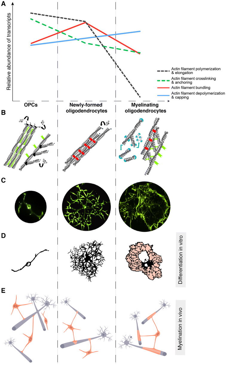
The organization of actin filaments into a wide range of subcellular structures is a defining feature of cell shape and dynamics, important for tissue development and homeostasis. Nervous system function requires morphological and functional plasticity of neurons and glial cells, which is largely determined by the dynamic reorganization of the actin cytoskeleton in response to intrinsic and extracellular signals. Oligodendrocytes are specialized glia that extend multiple actin-based protrusions to form the multilayered myelin membrane that spirally wraps around axons, increasing conduction speed and promoting long-term axonal integrity. Myelination is a remarkable biological paradigm in development, and maintenance of myelin is essential for a healthy adult nervous system. In this review, we discuss how structure and dynamics of the actin cytoskeleton is a defining feature of myelinating oligodendrocytes' biology and function. We also review "old and new" concepts to reflect on the potential role of the cytoskeleton in balancing life and death of myelin membranes and oligodendrocytes in the aging central nervous system.
Keywords: Age-associated cognitive decline; Brain aging; Cellular aging; Glia; Membrane remodeling; Myelin; White matter.
Figures
Similar articles
-
The Actin Cytoskeleton in Myelinating Cells.Neurochem Res. 2020 Mar;45(3):684-693. doi: 10.1007/s11064-019-02753-0. Epub 2019 Mar 7. Neurochem Res. 2020. PMID: 30847860 Free PMC article. Review.
-
Jmy regulates oligodendrocyte differentiation via modulation of actin cytoskeleton dynamics.Glia. 2018 Sep;66(9):1826-1844. doi: 10.1002/glia.23342. Epub 2018 May 6. Glia. 2018. PMID: 29732611
-
Daam2 Regulates Myelin Structure and the Oligodendrocyte Actin Cytoskeleton through Rac1 and Gelsolin.J Neurosci. 2022 Mar 2;42(9):1679-1691. doi: 10.1523/JNEUROSCI.1517-21.2022. Epub 2022 Jan 31. J Neurosci. 2022. PMID: 35101966 Free PMC article.
-
The cell biology of CNS myelination.Curr Opin Neurobiol. 2016 Aug;39:93-100. doi: 10.1016/j.conb.2016.04.013. Epub 2016 May 3. Curr Opin Neurobiol. 2016. PMID: 27152449 Free PMC article. Review.
-
N-Wasp Regulates Oligodendrocyte Myelination.J Neurosci. 2020 Aug 5;40(32):6103-6111. doi: 10.1523/JNEUROSCI.0912-20.2020. Epub 2020 Jun 29. J Neurosci. 2020. PMID: 32601246 Free PMC article.
Cited by
-
Actin polymerization state regulates osteogenic differentiation in human adipose-derived stem cells.Cell Mol Biol Lett. 2021 Apr 15;26(1):15. doi: 10.1186/s11658-021-00259-8. Cell Mol Biol Lett. 2021. PMID: 33858321 Free PMC article.
-
The oligodendrocyte growth cone and its actin cytoskeleton: A fundamental element for progenitor cell migration and CNS myelination.Glia. 2020 Jul;68(7):1329-1346. doi: 10.1002/glia.23735. Epub 2019 Nov 7. Glia. 2020. PMID: 31696982 Free PMC article. Review.
-
Method for Diagnosing Bearing Faults in Electromechanical Equipment Based on Improved Prototypical Networks.Sensors (Basel). 2023 May 4;23(9):4485. doi: 10.3390/s23094485. Sensors (Basel). 2023. PMID: 37177689 Free PMC article.
-
Cannabinoids modulate proliferation, differentiation, and migration signaling pathways in oligodendrocytes.Eur Arch Psychiatry Clin Neurosci. 2022 Oct;272(7):1311-1323. doi: 10.1007/s00406-022-01425-5. Epub 2022 May 27. Eur Arch Psychiatry Clin Neurosci. 2022. PMID: 35622101
-
The Effect of Meclofenoxate on the Transcriptome of Aging Brain of Nothobranchius guentheri Annual Killifish.Int J Mol Sci. 2022 Feb 24;23(5):2491. doi: 10.3390/ijms23052491. Int J Mol Sci. 2022. PMID: 35269638 Free PMC article.
References
Publication types
MeSH terms
Grants and funding
LinkOut - more resources
Full Text Sources
Medical
