Review
. 2019 Mar;39(2):517-560.
doi: 10.1002/med.21531. Epub 2018 Oct 9.

The multifaceted role of autophagy in cancer and the microenvironment

Affiliations
Review

The multifaceted role of autophagy in cancer and the microenvironment

Hendrik Folkerts et al. Med Res Rev. 2019 Mar.

Abstract

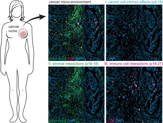
Autophagy is a crucial recycling process that is increasingly being recognized as an important factor in cancer initiation, cancer (stem) cell maintenance as well as the development of resistance to cancer therapy in both solid and hematological malignancies. Furthermore, it is being recognized that autophagy also plays a crucial and sometimes opposing role in the complex cancer microenvironment. For instance, autophagy in stromal cells such as fibroblasts contributes to tumorigenesis by generating and supplying nutrients to cancerous cells. Reversely, autophagy in immune cells appears to contribute to tumor-localized immune responses and among others regulates antigen presentation to and by immune cells. Autophagy also directly regulates T and natural killer cell activity and is required for mounting T-cell memory responses. Thus, within the tumor microenvironment autophagy has a multifaceted role that, depending on the context, may help drive tumorigenesis or may help to support anticancer immune responses. This multifaceted role should be taken into account when designing autophagy-based cancer therapeutics. In this review, we provide an overview of the diverse facets of autophagy in cancer cells and nonmalignant cells in the cancer microenvironment. Second, we will attempt to integrate and provide a unified view of how these various aspects can be therapeutically exploited for cancer therapy.

Keywords: autophagy; cancer; immune cells; microenvironment; stroma; therapy.


Figures

Figure 1
Figure 1
Review outline. This review highlights the impact of changes in autophagy within cancer cells, as well as in the context of the complex cancer microenvironment. Part I describes how aberrant autophagy can contribute to cancer initiation and maintenance as well as therapy resistance (pp. 6‐16). Part II describes the role of autophagy in different stromal cells within the tumor microenvironment, such as fibroblasts and mesenchymal stem cells (pp. 16‐18). Further, the impact of autophagy on anticancer immune responses is described (pp. 18‐27). Blue, 4′,6‐diamidino‐2‐phenylindole (DAPI) staining; green, fibronectin staining for stroma; red, CD8 staining for cytotoxic T‐cell [Color figure can be viewed at wileyonlinelibrary.com]
Figure 2
Figure 2
The autophagy pathway. A, The activation of autophagy is initiated by the reduced activity of the mechanistic target of rapamycin complex 1 (mTORC1) complex due to activated adenosine monophosphate‐activated protein kinase (AMPK) or decreased upstream growth signaling. mTORC1 is an inhibitor of the ULK complex, therefore reduced mTORC1 activity increases the activity of the ULK complex. The ULK complex together with the Beclin‐1/ VPS34 complex initiates the formation of autophagosomes. Dependent on the complex composition, Beclin‐1 can act as a molecular switch between autophagy and apoptosis (see B). The expansion and maturation of the autophagosomes is dependent on two ubiquitin‐like conjugation systems, which requires multiple autophagy proteins. First, ATG12‐ATG5 conjugate binds to ATG16, which stimulates LC3 lipidation. Second, LC3 is covalently conjugated to phosphatidylethanolamine (PE) generating LC3‐II, which is incorporated in the autophagosomal membrane. Incorporated LC3‐II is required for binding and internalization of adaptor proteins such as p62. Finally, the mature autophagosome fuses with lysosomes, after which its content is broken down by digestive enzymes. Indicated in red are pharmacological agents, chloroquine (CQ), hydroxychloroquine (HCQ), 3‐methyladenine (3‐MA), and ULK inhibitors, that inhibit autophagy. In addition, rapamycin activates autophagy by inhibiting mTORC1. B, Beclin‐1 is a core member of the VPS34/Beclin‐1 complex, which acts as a molecular switch in controlling autophagy downstream of the ULK1 complex. Depicted in red are the antiapoptotic members of the Bcl‐2 family BCL‐2, BCL‐XL, and MCL‐1 which can bind to Beclin‐1, through interaction with its BH3 domain, thereby inhibiting autophagy. Alternatively, Bcl‐2 interacting protein 3 (BNIP3) and Bcl‐2 interacting protein 3 like (BNIP3L; depicted in green) can competitively bind to antiapoptotic BLC‐2 members. Dissociation of antiapoptotic Bcl‐2 members from Beclin‐1, consequently activates autophagy. Other non‐BH3 proteins, also depicted in green, such as vacuole membrane protein 1 (VMP1), ATG14, UV radiation resistance‐associated gene (UVRAG), and activating molecule in Beclin‐1‐regulated autophagy protein 1 (AMBRA1) can also bind Beclin‐1, thereby activating autophagy. PDK1, pyruvate dehydrogenase kinase 1; PI3K, phosphoinositide 3‐kinase [Color figure can be viewed at wileyonlinelibrary.com]
Figure 3
Figure 3
Autophagy during malignant transformation and cancer maintenance. A, Different pro‐oncogenic events such as mutation or monoallelic deletion of autophagy‐related genes can cause reduced autophagy activity. Reduced levels of autophagy/mitophagy can contribute to malignant transformation due to elevated levels of reactive oxygen species (ROS). B, Hematopoietic stem cells (HSCs) reside in specific bone marrow niches with low oxygen content and are characterized by high autophagy activity. During differentiation, the autophagy flux declines and mature cells leave the bone marrow (BM) environment and enter the blood‐stream. In leukemia, HSCs have acquired mutations which results in a block in differentiation and consequently accumulation of immature blasts in BM and peripheral blood of patients. C, Hypothetical model for changes in autophagy and ROS in HSCs during transformation. Normal HSCs have high autophagy flux, low mitochondrial activity, and ROS levels. During cancer initiation, autophagy is repressed (although not completely inhibited), causing accumulation of mitochondria and ROS, which in turn contributes to malignant transformation. During cancer maintenance, cancer cells re‐establish functional autophagy promoting tumor growth and survival. In addition, in response to drug treatment, autophagy is activated and acts as a survival mechanism for cancer cells. D, Both normal BM‐derived CD34+ and acute myeloid leukemia (AML) CD34+ cells need a certain level of autophagy to survive. Therefore, there is only a small therapeutic window of autophagy inhibition with autophagy inhibitors like hydroxychloroquine. LSC, leukemic stem cells [Color figure can be viewed at wileyonlinelibrary.com]
Figure 4
Figure 4
Autophagy in the tumor microenvironment impacts on anticancer immunity. Autophagy in cancer cells inhibits the anticancer immune response by reducing the efficacy of cytotoxic T‐cell and natural killer cell–mediated lysis by degrading granzyme B and connexin‐43. Further, autophagy is also required for T‐cell proliferation, survival, and induction of T‐cell memory by degrading proapoptotic proteins and maintaining mitochondrial homeostasis. Therefore, nonselective inhibition of autophagy in the tumor microenvironment will not only promote anticancer effects at the level of stroma and cancer cells, but will also dampen anticancer immune responses. CDKN1B, cyclin‐dependent kinase inhibitor 1B; PD‐L1, programmed cell death 1 and its ligand; TCR, T‐cell receptor [Color figure can be viewed at wileyonlinelibrary.com]
Figure 5
Figure 5
Autophagy contributes to the formation of antigenic peptides in antigen‐presenting cells (APCs). Professional APCs, such as dendritic cells and macrophages, display antigenic peptides in the context of major histocompatibility complex class 1 (MHC‐I) or MHC‐II molecules to T‐cells, which will trigger an immune response. Autophagy reduces MHC‐I surface levels, which is converted upon autophagy inhibition. However, autophagy is also required for the generation of antigenic peptides. The inhibition of autophagy will therefore skew the peptidome, yielding less diversity In the antigens presented to T‐cells. Indeed autophagy, inhibition limits T‐cell activation by APCs [Color figure can be viewed at wileyonlinelibrary.com]

References

    1. Kroemer G, Mariño G, Levine B. Autophagy and the integrated stress response. Mol Cell. 2010;40(2):280‐293. - PMC - PubMed
    1. Kubli DA, Gustafsson AB. Mitochondria and mitophagy: the yin and yang of cell death control. Circ Res. 2012;111(9):1208‐1221. - PMC - PubMed
    1. Ding W‐X, Yin X‐M. Mitophagy: mechanisms, pathophysiological roles, and analysis. Biol Chem. 2012;393(7):547‐564. - PMC - PubMed
    1. White E. The role for autophagy in cancer. J Clin Invest. 2015;125(1):42‐46. - PMC - PubMed
    1. Allen GFG, Toth R, James J, Ganley IG. Loss of iron triggers PINK1/Parkin‐independent mitophagy. EMBO Rep. 2013;14:1127‐1135. - PMC - PubMed

Publication types

MeSH terms

LinkOut - more resources