

Focal Degeneration of Vestibular Neuroepithelium in the Cristae Ampullares of Three Human Subjects
- PMID: 30303940
- PMCID: PMC6242718
- DOI: 10.1097/MAO.0000000000002018
Focal Degeneration of Vestibular Neuroepithelium in the Cristae Ampullares of Three Human Subjects
Abstract
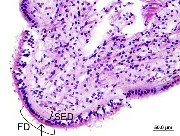
Background: We report a unique pattern of focal degeneration of the neuroepithelium of cristae ampullares, thick subepithelial extracellular deposits, and neural degeneration in three humans.
Objective: To characterize the pattern of vestibular degeneration and measure the thickness of subepithelial deposits in these three cases and controls.
Methods: The subepithelial deposits of vestibular end organs in three subject cases and controls were studied using hematoxylin and eosin, periotic acid-Schiff, Gomori trichrome staining, and immunostaining for antineurofilament, antimyosin VIIa, and anticollagen 4a1. The thickness of deposit as measured by light microscopy was compared with that of control groups (age-matched controls, patients with unilateral Menière's disease, vestibular neuritis, cupulolithiasis, severe nonfocal degeneration of the vestibular neuroepithelium, and Alport syndrome). The correlation of thickness of deposits with age from 0 to 100 years was also investigated.
Results: Focal loss of hair cells in the neuroepithelium, thick subepithelial deposits, and degeneration of subepithelial dendrites and Scarpa's ganglion were found in all three cristae of three subject cases. Immunostaining demonstrated a decrease of afferent neural fibers in the cristae and focal fragmentation of the basement membrane adjacent to the deposits. The thickness of the subepithelial deposits in three cristae of three subject cases was significantly greater than that of all controls. In the three cristae of normal controls, the thickness of deposits demonstrated a positive correlation with age.
Conclusion: Although both age and degeneration of the vestibular neuroepithelium may be associated with the thickness of the subepithelial deposits, in this unique pattern of degeneration, the thickness of the subepithelial deposits was significantly greater than that in all controls.
Conflict of interest statement
(disclosure)
All author report no conflict of interest related to this manuscript.
Figures


References
-
- Wersall J Studies on the structure and innervation of the sensory epithelium of the cristae ampulares in the guinea pig; a light and electron microscopic investigation. Acta Otolaryngol Suppl 1956;126:1–85. - PubMed
-
- Merchant SN, Velazquez-Villaseňor L, Tsuji K, Glynn RJ, Wall C 3rd, Rauch SD. Temporal bone studies of the human peripheral vestibular system. Normative vestibular hair cell data. Ann Otol Rhinol Suppl 2000;181:3–13. - PubMed
-
- Merchant SN, Burgess BJ, Adams JC, et al. Temporal bone histopathology in Alport syndrome. Laryngoscope 2004;114:1609–18. - PubMed
Publication types
MeSH terms
Grants and funding
LinkOut - more resources
Full Text Sources
