

doi: 10.3390/molecules23102572.
2'- O-Methyl-8-methylguanosine as a Z-Form RNA Stabilizer for Structural and Functional Study of Z-RNA
Affiliations
- PMID: 30304782
- PMCID: PMC6222775
- DOI: 10.3390/molecules23102572
Item in Clipboard
2'- O-Methyl-8-methylguanosine as a Z-Form RNA Stabilizer for Structural and Functional Study of Z-RNA
Molecules.
.
Abstract
In contrast to Z-DNA that was stabilized and well-studied for its structure by chemical approaches, the stabilization and structural study of Z-RNA remains a challenge. In this study, we developed a Z-form RNA stabilizer m⁸Gm, and demonstrated that incorporation of m⁸Gm into RNA can markedly stabilize the Z-RNA at low salt conditions. Using the m⁸Gm-contained Z-RNA, we determined the structure of Z-RNA and investigated the interaction of protein and Z-RNA.
Keywords: NMR; Z-RNA structure; circular dichroism; synthesis of oligonucleotide.
Conflict of interest statement
The authors declare no conflict of interest.
Figures
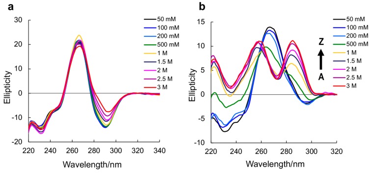
CD spectra of native RNA r(CGCGCG)2 (a) and m8Gm-contained r(CGC[m8Gm]CG)2 (b) (0.15 mM base concentration) at 10 °C with various NaClO4 concentrations in 5 mM sodium phosphate buffer (pH 7.0).
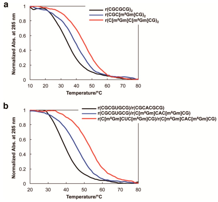
(a) CD melting curves for native RNA r(CGCGCG)2 (back line), r(CGC[m8Gm]CG)2 containing one m8Gm (blue line), and r(C[m8Gm]C[m8Gm]CG)2 containing two m8Gms (red line). (b) CD melting curves for RNA sequences containing an AU base pair, native RNA r(CGCGUGCG)/r(CGCACGCG) (back line), r(CGCGUGCG)/r(C[m8Gm]CAC[m8Gm]CG) having two m8Gms in one strand (blue line), r(C[m8Gm]CGU[m8Gm]CG)r(C[m8Gm]CAC[m8Gm]CG) having four m8Gms in two strands (red line). In 5 mM sodium phosphate buffer (pH 7.0), 7 M NaClO4 concentrations (0.15 mM base concentration).
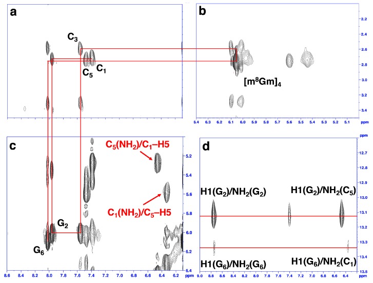
(a–c) H6/H5′′ of C and H8/H1′ of G (8CH3/H1′ of m8Gm) proton region of NOESY spectra of r(CGC[m8Gm]CG)2 in NaClO4 solution. The NOE connectivity pathway is shown as red line. Intraresidue NOE cross-peaks are labeled with residue numbers. The Z-RNA structure specific cross-peaks were observed between C5 amino protons and C1H5 protons from the opposite strand (indicated as red words), as well as between C1 amino protons and C5H5 protons from the opposite strand (c). (d) The cross peaks of imino proton of G2 and amino proton of C5, as well as imino proton of G6 and amino proton of C1, indicated Watson–Crick-type base pairs of Z-RNA. Intraresidue NOE cross-peaks of imino and amino proton of G2 and G6 are shown.
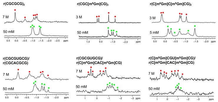
Monitoring of the A to Z-RNA transition by 31P NMR. 31P NMR spectra of the RNAs at 5 mM-7 M NaClO4. Green and red dots indicate the 31P peaks resulting from A-RNA and Z-RNA, respectively.
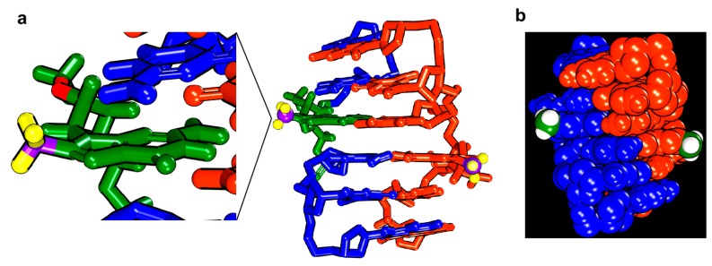
The model for r(CGC[m8Gm]CG)2 Z-RNA structure. (a) m8Gm is expanded in green with the C8-methyl group, hydrogen and carbon of methyl group are coloured in yellow and purple. (b) Hydrogen and carbon of methyl group are coloured in green and white. C8-methyl groups were located outside of Z-RNA.

Visualization of Z-RNA and Zα-EGFP protein. Lane 1: Z-RNA only, Lane 2: RNA + Zα-EGFP, Lane 3: Zα-EGFP only. The different modes are indicated in upper. [RNA] = 1 μM, [Zα-EGFP] = 10 μM, [NaCl] = 100 mM, [Tris-HCl (pH7.0)] = 10 mM, [DTT] = 5 mM, 10% glycerol, 10 μg/mL BSA. Z-RNA: r(CGCGUGCG)-Cy3/r(C[m8Gm]CAC[m8Gm]CG).
References
MeSH terms
Substances
Grants and funding
LinkOut - more resources
Full Text Sources
