. 2019 Sep;37(15):3968-3975.
doi: 10.1080/07391102.2018.1532817. Epub 2019 Jan 11.

Crystal structure of a dimerization domain of human Caprin-2: similar overall dimeric fold but different molecular surface properties to that of human Caprin-1

Affiliations

Crystal structure of a dimerization domain of human Caprin-2: similar overall dimeric fold but different molecular surface properties to that of human Caprin-1

Yuhong Wu et al. J Biomol Struct Dyn. 2019 Sep.

Abstract

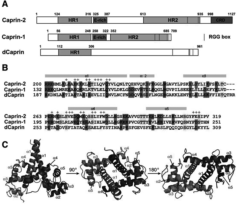
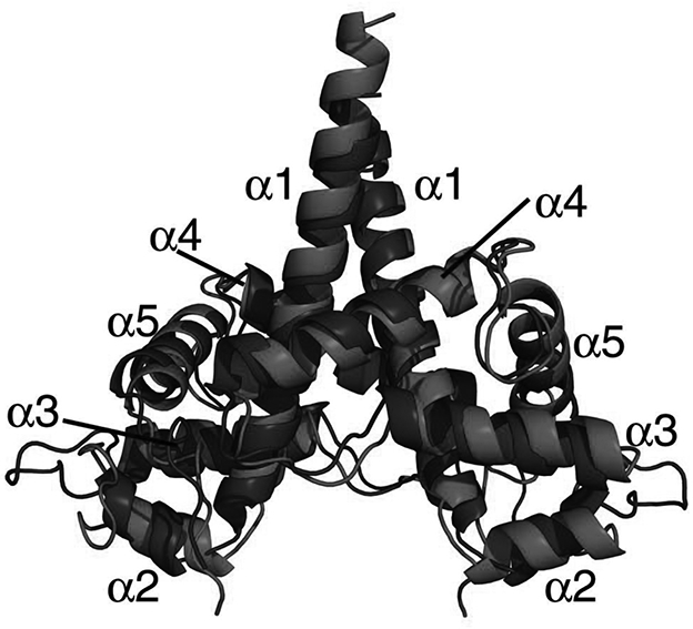
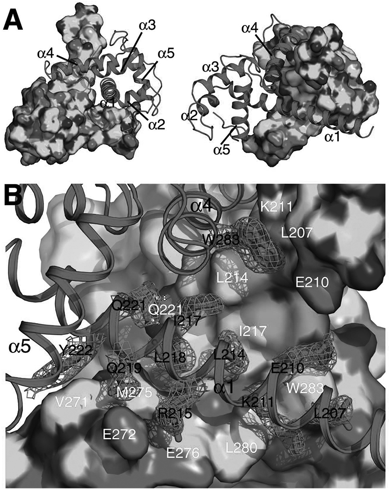
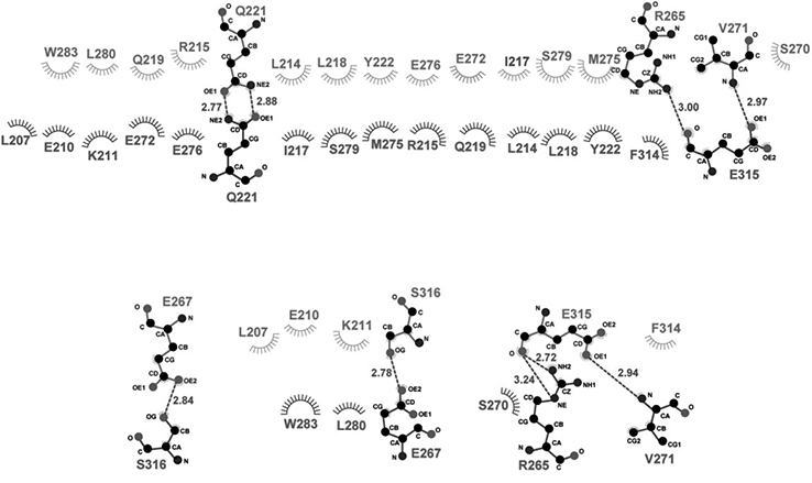
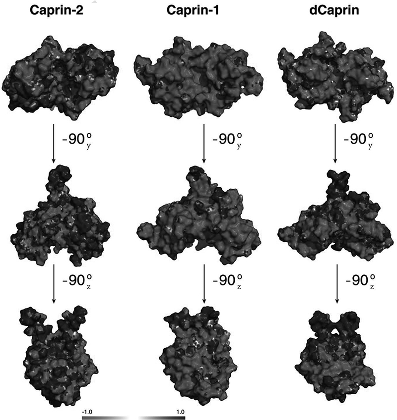
Human Caprin-1 and Caprin-2 are prototypic members of the caprin (cytoplasmic activation/proliferation-associated protein) protein family. Vertebrate caprin proteins contain two highly conserved homologous regions (HR1 and HR2) and C-terminal RGG motifs. Drosophila caprin (dCaprin) shares HR1 and RGG motifs but lacks HR2. Caprin-1 and Caprin-2 have important and non-redundant functions. The detailed molecular mechanisms of their actions remain largely unknown. Previously, we determined the crystal structure of a ∼120-residue fragment of Caprin-1 within the HR1 region. The structure has a novel all α-helical fold that self-associates to form a homodimer. In this study, the crystal structure of a corresponding fragment from Caprin-2 is reported. The Caprin-2 fragment has similar protein fold and dimeric structure as that of the Caprin-1 fragment. Structural comparison reveals that the molecular interactions mediating homodimerization of Caprin-1 and Caprin-2 are largely conserved in the two systems. Structural-modelling study of the corresponding dCaprin fragment indicates that dCaprin may also adopt a similar dimeric structure. The presence of a dimerization domain within HR1 may represent an evolutionarily conserved feature of the caprin protein family. Interestingly, while Caprin-1 and Caprin-2 adopt similar overall dimeric structures, the two structures have quite different molecular surface properties. In the Caprin-1 dimeric structure, some of the surface areas are known or suspected to function as binding sites for Carpin-1-interacting proteins. The different surface properties of the caprin dimeric structures may dictate their intermolecular interaction with specific protein partners. Communicated by Ramaswamy H. Sarma.

Keywords: Caprin-1; FMRP; G3BP1; RNA stress granule; caprin-2.


Figures

Figure 1.
Figure 1.. Caprin proteins and structure of the dimerization domain of Caprin-2.
A) Schematic representation of human Caprin-2 and Caprin-1, as well as Drosoplila Caprin (dCaprin). HR1 and HR2: homologous region 1 and 2. CRD: C1q-related domain. The thin red lines indicate the locations of the RGG boxes. B) Sequence of the human Caprin-2 dimerization domain aligned with homologous sequences of Caprin-1 and dCaprin. The secondary structures are indicated above the Caprin-2 sequence. Residues involved in Caprin-2 dimerization are indicate with “+” sign above the sequence. C) Structure of the Caprin-2 homodimer rendered in cartoon mode, viewed from three different angles. The two protomers are coloured red and blue respectively.
Figure 2.
Figure 2.
Superimposition of the dimeric structures of the Caprin-2 (in red, PDB code 5J97) and Caprin-1 (in blue, PDB code 4WBE).
Figure 3.
Figure 3.. Molecular interactions that mediate the homodimerization of Caprin-2.
A) Shape complementarity at the homodimerization interface. The two protomers are rendered in surface mode (coloured by elements with C, N, O and S in green, blue, red and yellow respectively) and cartoon mode (coloured in magenta) respectively. B) Intermolecular hydrophobic contacts, salt bridges, and hydrogen bonds at the homodimerization interface. The side-chains for some of the key residues belonging to the protomer rendered in cartoon mode are shown in sticks. 2Fo-Fc electron density map (mesh) contoured at 1σ is shown for these side-chains. The residues involved in the intermolecular interactions are indicated differently for the two protomers: one letter abbreviation-residue number in black for residues belonging to the protomer rendered in cartoon mode and one letter abbreviation-residue number in white for residues belonging to the protomer rendered in surface mode. Hydrogen bonds are represented as yellow dashed lines.
Figure 4.
Figure 4.. Ligplot presentation of molecular interactions in the dimerization interface.
Hydrogen bonds are designated with green dashed lines (the distance between the two heavy atoms is indicated by a number in angstrom). Hydrophobic interactions are represented as starbursts.
Figure 5.
Figure 5.. Electrostatic surface representation of Caprin homodimeric structures (for Caprin-2 and Caprin-1) and a structural model (for dCaprin).
Blue and red represent regions of positive and negative potentials respectively.

References

    1. Adams PD, Afonine PV, Bunkoczi G, Chen VB, Echols N, Headd JJ, … Zwart PH (2011). The Phenix software for automated determination of macromolecular structures. Methods, 55(1), pp. 94–106. doi:10.1016/j.ymeth.2011.07.005 - DOI - PMC - PubMed
    1. Aerbajinai W, Lee YT, Wojda U, Barr VA, & Miller JL (2004). Cloning and characterization of a gene expressed during terminal differentiation that encodes a novel inhibitor of growth. J Biol Chem, 279(3), pp. 1916–1921. doi:10.1074/jbc.M305634200 - DOI - PubMed
    1. Afonine PV, Grosse-Kunstleve RW, Echols N, Headd JJ, Moriarty NW, Mustyakimov M, … Adams PD (2012). Towards automated crystallographic structure refinement with phenix.refine. Acta Crystallogr D Biol Crystallogr, 68(Pt 4), pp. 352–367. doi:10.1107/s0907444912001308 - DOI - PMC - PubMed
    1. Battye TG, Kontogiannis L, Johnson O, Powell HR, & Leslie AG (2011). iMOSFLM: a new graphical interface for diffraction-image processing with MOSFLM. Acta Crystallogr D Biol Crystallogr, 67(Pt 4), pp. 271–281. doi:10.1107/s0907444910048675 - DOI - PMC - PubMed
    1. Biasini M, Bienert S, Waterhouse A, Arnold K, Studer G, Schmidt T, … Schwede T (2014). SWISS-MODEL: modelling protein tertiary and quaternary structure using evolutionary information. Nucleic Acids Res, 42(Web Server issue), pp. W252–258. doi:10.1093/nar/gku340 - DOI - PMC - PubMed

Publication types

LinkOut - more resources