. 2018 Oct 11;9(1):265.
doi: 10.1186/s13287-018-1007-x.

Short-term physiological hypoxia potentiates the therapeutic function of mesenchymal stem cells

Affiliations

Short-term physiological hypoxia potentiates the therapeutic function of mesenchymal stem cells

Ben Antebi et al. Stem Cell Res Ther. .

Abstract

Background: In the bone marrow, MSCs reside in a hypoxic milieu (1-5% O2) that is thought to preserve their multipotent state. Typically, in vitro expansion of MSCs is performed under normoxia (~ 21% O2), a process that has been shown to impair their function. Here, we evaluated the characteristics and function of MSCs cultured under hypoxia and hypothesized that, when compared to normoxia, dedicated hypoxia will augment the functional characteristics of MSCs.

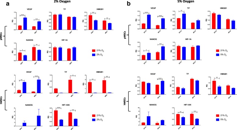
Methods: Human and porcine bone marrow MSCs were obtained from fresh mononuclear cells. The first study evaluated MSC function following both long-term (10 days) and short-term (48 h) hypoxia (1% O2) culture. In our second study, we evaluated the functional characteristics of MSC cultured under short-term 2% and 5% hypoxia. MSCs were evaluated for their metabolic activity, proliferation, viability, clonogenicity, gene expression, and secretory capacity.

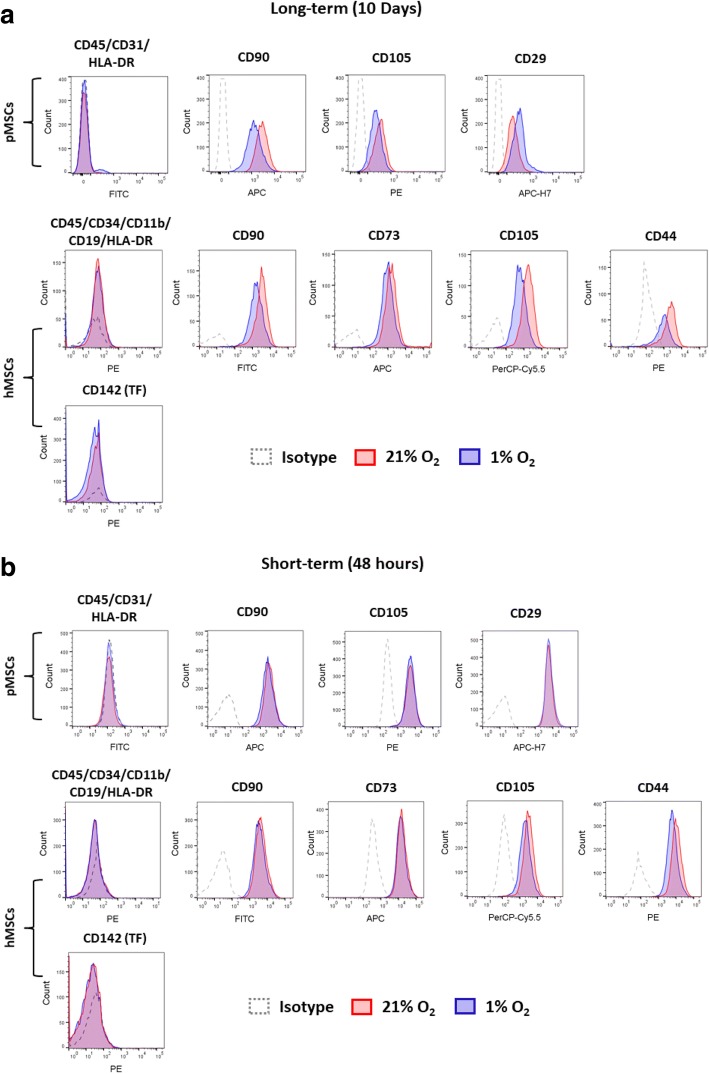
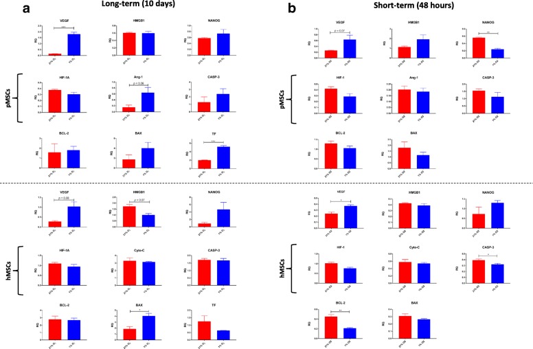
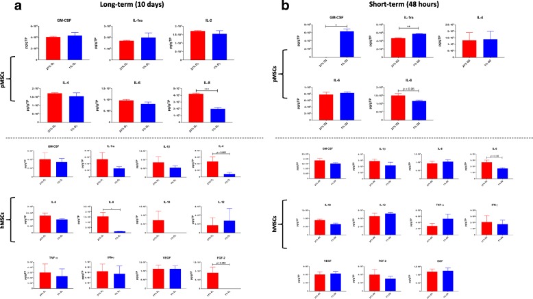
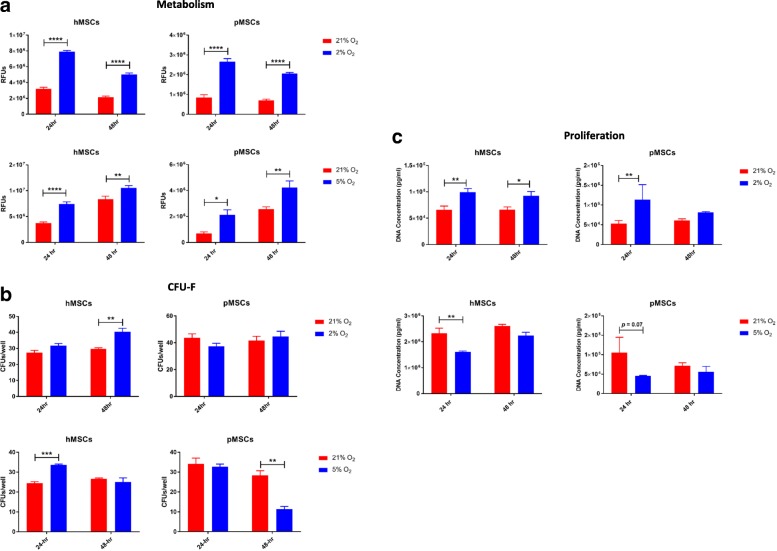
Results: In long-term culture, common MSC surface marker expression (CD44 and CD105) dropped under hypoxia. Additionally, in long-term culture, MSCs proliferated significantly slower and provided lower yields under hypoxia. Conversely, in short-term culture, MSCs proliferated significantly faster under hypoxia. In both long-term and short-term cultures, MSC metabolic activity was significantly higher under hypoxia. Furthermore, MSCs cultured under hypoxia had upregulated expression of VEGF with concomitant downregulation of HMGB1 and the apoptotic genes BCL-2 and CASP3. Finally, in both hypoxia cultures, the pro-inflammatory cytokine, IL-8, was suppressed, while levels of the anti-inflammatories, IL-1ra and GM-CSF, were elevated in short-term hypoxia only.

Conclusions: In this study, we demonstrate that hypoxia augments the therapeutic characteristics of both porcine and human MSCs. Yet, short-term 2% hypoxia offers the greatest benefit overall, exemplified by the increase in proliferation, self-renewing capacity, and modulation of key genes and the inflammatory milieu as compared to normoxia. These data are important for generating robust MSCs with augmented function for clinical applications.

Keywords: Bone marrow; Hypoxia; Immunomodulation; Mesenchymal stem cells (MSCs); Normoxia.


Conflict of interest statement

Ethics approval and consent to participate

N/A

Consent for publication

N/A

Competing interests

The authors declare that they have no competing interests.

Publisher’s Note

Springer Nature remains neutral with regard to jurisdictional claims in published maps and institutional affiliations.

Figures

Fig. 1
Fig. 1
Surface expression of MSC markers under long-term and short-term hypoxia. (a) In long-term hypoxia, CD44 levels are decreased in hMSCs, especially under hypoxia. Levels of CD105 are decreased under hypoxia, in both human and porcine MSCs. No expression of TF (CD142) was evident in hMSCs under long-term normoxia or hypoxia culture. (b) In short-term hypoxia, all surface markers are nearly 100% besides CD105, which is decreased in hMSCs. No expression of TF was evident in hMSCs under short-term normoxia or hypoxia culture
Fig. 2
Fig. 2
Characteristics of MSCs under long-term and short-term hypoxia. a Compared to normoxia, the metabolic activity of MSCs is significantly increased under 1% oxygen, particularly in long-term cultures; b In short-term culture, pMSCs show significant increase in self-renewing capacity as compared to normoxia (p < 0.01); c MSCs proliferate faster and generate greater cell yields under normoxia in long-term culture, while in short-term culture, MSCs demonstrate enhanced proliferation capacity under hypoxia; d In long-term culture, viable MSCs, shown in fluorescent green, reach a confluent state faster under normoxia; bar is 500 μm; *p < 0.05; **p < 0.01; ***p < 0.001; ****p < 0.0001
Fig. 3
Fig. 3
Gene expression of MSCs under long-term and short-term hypoxia. a In long-term cultures, TF, VEGF, and Ang-1 genes are upregulated in pMSCs under hypoxia. In hMSCs, VEGF (p = 0.08) and BAX (p < 0.05) were upregulated under hypoxia while HMGB1 (p = 0.08) was downregulated. (b) In short-term hypoxia culture, VEGF (p = 0.07) was upregulated while NANOG (p < 0.01) was downregulated in pMSCs. In hMSCs, VEGF (p < 0.05) was upregulated under hypoxia while the pro-apoptotic genes, BCL-2 (p < 0.01) and CASP-3 (p < 0.05), were downregulated. *p < 0.05; **p < 0.01; ***p < 0.001
Fig. 4
Fig. 4
Secretion profile of MSCs under long-term and short-term hypoxia. (a) In long-term culture, IL-8 levels were suppressed under hypoxia in both human and porcine MSCs. (b) In short-term culture, IL-8 was similarly suppressed under hypoxia in both human and porcine MSCs (p < 0.06). In pMSCs, significantly higher levels of the anti-inflammatories, GM-CSF (p < 0.05) and IL-1ra (p < 0.01) were secreted under hypoxia, as compared to normoxia
Fig. 5
Fig. 5
Surface expression of MSC markers under 2% and 5% short-term hypoxia. (a) Under 2% hypoxia, all MSC positive surface markers were above 95%. (b) In contrast, under 5% hypoxia, there was a significant reduction (p < 0.0001) in CD105 expression in hMSCs at noth 24 and 48 hours. This reduction was not evident in pMSCs where surface expression was maintained above 99%
Fig. 6
Fig. 6
MSC characteristics under 2% and 5% short-term hypoxia. a The metabolic activity of MSCs was significantly elevated under both 2% and 5% hypoxia, when compared to normoxia; b The clonogenic capacity of hMSCs was significantly increased under hypoxia, while for pMSCs it was decreased after 48 h under 5% hypoxia; c Proliferation of MSCs was significantly increased under 2% hypoxia, whereas a significantly lower rate of proliferation was seen under 5% hypoxia; *p < 0.05; **p < 0.01; ***p < 0.001; ****p < 0.0001
Fig. 7
Fig. 7
Gene expression under 2% and 5% short-term hypoxia. (a) Under 2% short-term hypoxia, there was a significant upregulation in the angiogenic gene, VEGF, with a concomitant downregulation in the expression of HMBG1 in both human and porcine MSCs. Additionally, under hypoxia, the stem cell gene, NANOG, was downregulated in porcine, but not human MSCs, while TF and HIF antagonist were downregulated in human, but not porcine MSCs. (b) Similar to 2% hypoxia, under 5% hypoxia, the VEGF gene was significantly upregulated under hypoxia in both human and porcine MSCs, while HMGB1 and HIF antagonist were downregulated in hMSCs. As opposed to 2% hypoxia, TF was significantly upregulated in pMSCs under 5% hypoxia; *p < 0.05; **p < 0.01; ***p < 0.001; ****p < 0.0001
Fig. 8
Fig. 8
Secretion profile of MSCs under 2% and 5% short-term hypoxia. (a) Under 2% short-term hypoxia, IL-6 and IL-8 secreted by pMSCs were significantly suppressed while GM-CSF, IFN-γ, IL-1ra, and IL-18 were significantly increased. In hMSCs, IFN-γ and IL-18 were significantly increased under 2% hypoxia. (b) Similar to 2% hypoxia, 5% hypoxia significantly suppressed the secretion of the pro-inflammatories IL-6 and IL-8 in pMSCs. Unlike 2%, 5% hypoxia concomitantly suppressed the secretion of GM-CSF, IFN-γ, IL-1α, IL-2, IL-4, and IL-10. In hMSCs, as in pMSCs, IL-8 and GM-CSF levels were suppressed, while the secretion of IL-12 was increased under hypoxia, similar to the 2% hypoxia; *p < 0.05; **p < 0.01; ***p < 0.001; ****p < 0.0001

References

    1. Weiss DJ. Concise review: current status of stem cells and regenerative medicine in lung biology and diseases. Stem Cells. 2014;32(1):16–25. doi: 10.1002/stem.1506. - DOI - PMC - PubMed
    1. Lee JW, Fang X, Krasnodembskaya A, Howard JP, Matthay MA. Concise review: Mesenchymal stem cells for acute lung injury: role of paracrine soluble factors. Stem Cells. 2011;29(6):913–919. doi: 10.1002/stem.643. - DOI - PMC - PubMed
    1. Antebi Ben, Mohammadipoor Arezoo, Batchinsky Andriy I., Cancio Leopoldo C. The promise of mesenchymal stem cell therapy for acute respiratory distress syndrome. Journal of Trauma and Acute Care Surgery. 2018;84(1):183–191. doi: 10.1097/TA.0000000000001713. - DOI - PubMed
    1. Matthay MA, Thompson BT, Read EJ, Jr. McKenna DH, Liu KD, Calfee CS, Lee JW. Therapeutic potential of mesenchymal stem cells for severe acute lung injury. Chest. 2010;138(4):965–972. doi: 10.1378/chest.10-0518. - DOI - PMC - PubMed
    1. Gupta N, Su X, Popov B, Lee JW, Serikov V, Matthay MA. Intrapulmonary delivery of bone marrow-derived mesenchymal stem cells improves survival and attenuates endotoxin-induced acute lung injury in mice. J Immunol. 2007;179(3):1855–1863. doi: 10.4049/jimmunol.179.3.1855. - DOI - PubMed

Publication types

MeSH terms