. 2018 Oct 23;115(43):10983-10988.
doi: 10.1073/pnas.1802932115. Epub 2018 Oct 10.

SUMO protease SENP1 deSUMOylates and stabilizes c-Myc

Affiliations

SUMO protease SENP1 deSUMOylates and stabilizes c-Myc

Xiao-Xin Sun et al. Proc Natl Acad Sci U S A. .

Abstract

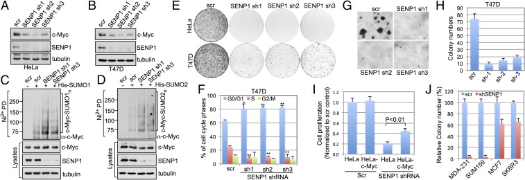
Posttranslational modifications play a crucial role in the proper control of c-Myc protein stability and activity. c-Myc can be modified by small ubiquitin-like modifier (SUMO). However, how SUMOylation regulates c-Myc stability and activity remains to be elucidated. The deSUMOylation enzyme, SENP1, has recently been shown to have a prooncogenic role in cancer; however, mechanistic understanding of this is limited. Here we show that SENP1 is a c-Myc deSUMOylating enzyme. SENP1 interacts with and deSUMOylates c-Myc in cells and in vitro. Overexpression of wild-type SENP1, but not its catalytically inactive C603S mutant, markedly stabilizes c-Myc and increases its levels and activity. Knockdown of SENP1 reduces c-Myc levels, induces cell cycle arrest, and drastically suppresses cell proliferation. We further show that c-Myc can be comodified by both ubiquitination and SUMOylation. SENP1-mediated deSUMOylation reduces c-Myc polyubiquitination, suggesting that SUMOylation promotes c-Myc degradation through the proteasome system. Interestingly, SENP1-mediated deSUMOylation promotes the accumulation of monoubiquitinated c-Myc and its phosphorylation at serine 62 and threonine 58. SENP1 is frequently overexpressed, correlating with the high expression of c-Myc, in breast cancer tissues. Together, these results reveal that SENP1 is a crucial c-Myc deSUMOylating enzyme that positively regulates c-Myc's stability and activity.

Keywords: SENP1; SUMOylation; c-Myc; deSUMOylation; ubiquitination.


Conflict of interest statement

The authors declare no conflict of interest.

Figures

Fig. 1.
Fig. 1.
SENP1 interacts with c-Myc in cells and in vitro. (A and B) Co-IP of ectopic SENP1 with ectopic Myc. H1299 cells transfected with V5–c-Myc and Flag-SENP1 (A) or V5-SENP1 and Flag–c-Myc (B) individually or together were assayed by co-IP. (C) Co-IP between endogenous SENP1 and c-Myc using 293 cell lysates. (D) SENP1 interacts with c-Myc in vitro. Purified GST or GST-SENP1 immobilized on glutathione beads was incubated with purified His-Myc. Bound proteins were assayed by IB with anti-Myc (Top). Coomassie staining of GST and GST-SENP1 proteins is shown (Bottom). (E) HeLa cells were stained with anti–c-Myc and anti-SENP1 antibodies followed by PLA. The PLA signals were overlaid with DAPI from confocal imaging.
Fig. 2.
Fig. 2.
SENP1 deSUMOylates and stabilizes c-Myc and increases its activity. (A and B) SENP1 deSUMOylates c-Myc in cells. H1299 cells were transfected with His-SUMO1 (A) or His-SUMO2 (B) with or without Flag-SENP1 (WT or the C603S mutant) for 48 h and treated with MG132 for 6 h. The cells were subjected to pulldown (PD) using Ni2+-NTA bead under denaturing conditions, followed by IB. (C) SENP1 deSUMOylates c-Myc in vitro. SUMOylated c-Myc purified from 293 cells transfected with Flag-SUMO2 and c-Myc was incubated with recombinant WT GST-SENP1 or its C603S mutant, followed by IB. (D and E) SENP1 induces c-Myc levels in cells. HeLa cells transfected with increasing amounts of WT Flag-SENP1 (D) or with the C603S mutant (E), followed by IB. (F) SENP1 stabilizes c-Myc. HeLa cells transfected with control, WT SENP1, or its C603S mutant plasmid for 48 h were treated with 50 μg/mL cycloheximide (CHX). The cells were harvested at different time points and assayed by IB. (G) SENP1 increases c-Myc transactivation activity. H1299 cells transfected with luciferase reporter containing the E-box element from the E2F2 promoter in the presence of c-Myc and WT SENP1 or the C603S mutant, followed by luciferase activity measurement. *P < 0.05, compared with c-Myc only. (H) SENP1 stimulates c-Myc activity. U2OS-TO-Flag-SENP1WT or U2OS-TO-Flag-SENP1C603S cells were cultured in the absence or presence of doxycycline and assayed for gene expression by RT-qPCR. *P < 0.05, **P < 0.01, compared with the absence of doxycycline.
Fig. 3.
Fig. 3.
SENP1 inhibits c-Myc polyubiquitination and degradation. (A) Proteasome inhibitor treatment leads to accumulation of SUMOylated c-Myc with SUMO1 and SUMO2. HeLa cells transfected with His-SUMO1 or His-SUMO2 were treated with MG132 or DMSO for 6 h. The cells were subjected to Ni2+-NTA pulldown (PD) under denaturing conditions followed by IB. The SUMOylated c-Myc species are indicated. (B and C) SENP1 reduces polyubiquitination of c-Myc, but increases monoubiquitinated c-Myc. HeLa cells transfected with His-Ub and Flag-SENP1 as indicated and treated with MG132 (B) or without MG132 (C). The cells were subjected to Ni2+-NTA PD followed by IB. The polyubiquitinated (B) and monoubiquitinated (C) c-Myc species are indicated. (D) Co-IP of SENP1 with Fbw7α in H1299 cells transfected with V5-SENP1 with or without Flag-Fbw7α using anti-Flag antibody. (E) SENP1 inhibits c-Myc degradation mediated by Fbw7α. HeLa cells transfected with the indicated plasmids were assayed by IB. (F) SENP1 induces c-Myc levels in Fbw7+/+, but not Fbw7−/−, HCT116 cells. Cells transfected with control or Flag-SENP1 were assayed by IB. (G) Knockdown of SENP1 reduces c-Myc levels in Fbw7+/+, but not Fbw7−/−, HCT116 cells. The cells were infected with scrambled (scr) or SENP1 shRNA lentiviruses followed by IB. (H) SENP1 inhibits Fbw7-mediated ubiquitination of c-Myc. HeLa cells transfected with the indicated plasmids were treated with MG132 before harvesting. The cells were subjected to Ni2+-NTA PD under denaturing conditions followed by IB.
Fig. 4.
Fig. 4.
SENP1 induces c-Myc phosphorylation. (A) Overexpression of WT SENP1, but not the C603S mutant, increases c-Myc phosphorylation. HeLa cells transfected with the indicated plasmids were assayed by IB. Quantification is shown in SI Appendix, Fig. S6A. (B) Knockdown of SENP1 reduces c-Myc phosphorylation. HeLa cells infected with scrambled or SENP1 shRNA lentiviruses were assayed by IB. Quantification is shown in SI Appendix, Fig. S6B. (C) SENP1 is associated with chromatin. Nuclei isolated from HeLa cells transfected with the indicated plasmids were fractionated to chromatin and soluble fractions, followed by IB. (D) Binding of SENP1 with pS62–c-Myc in cells determined by PLA. HeLa cells were stained with anti-SENP1 and anti–pS62-Myc antibodies followed by PLA. The PLA signals are shown in the nucleus with enrichment in the nuclear periphery.
Fig. 5.
Fig. 5.
Knockdown of SENP1 reduces c-Myc levels and suppresses cell proliferation. (A and B) Knockdown of SENP1 reduces c-Myc levels. HeLa (A) or T47D (B) cells were infected with scrambled or SENP1 shRNA-encoding lentiviruses, followed by IB. (C and D) Knockdown of SENP1 increases c-Myc SUMOylation. H1299 cells transfected with His-SUMO1 (C) or His-SUMO2 (D) were infected with scrambled or SENP1 shRNA lentiviruses. The cells were assayed by Ni2+-NTA pulldown (PD) under denaturing conditions followed by IB. (E) Knockdown of SENP1 suppresses cell proliferation. HeLa or breast cancer T47D cells infected with control or SENP1 shRNA lentiviruses were cultured for up to 3 wk. The colonies were visualized by staining with crystal violet. (F) Knockdown of SENP1 induces G1 cell cycle arrest. T47D cells were infected with control or SENP1 shRNA lentiviruses followed by cell cycle analysis using propidium iodide staining. The mean percentage of cells in different cell cycle phases obtained from three independent experiments is shown. *P < 0.05, **P < 0.01, compared with the scrambled control. (G and H) Soft agar colony formation assays. T47D cells were infected with control or SENP1 shRNA lentiviruses, followed by colony formation assay in soft agar. Representative images are shown in G and average colony numbers were shown in H. (I) Overexpression of c-Myc partially alleviates the growth inhibition by SENP1 knockdown. Control or c-Myc–expressing HeLa cells were infected with scrambled or SENP1 shRNA lentiviruses, followed by MTT cell proliferation assays. (J) Knockdown of SENP1 inhibits anchorage-independent growth of Myc-sensitive breast cancer cells (SUM159 and MDA-MB-231) more efficiently than that of Myc-insensitive breast cancer cells (MCF7 and SKBR3). Shown is relative inhibition of colony formation in soft agar compared SENP1 shRNA infected cells to scramble control (J).
Fig. 6.
Fig. 6.
SENP1 is overexpressed in breast cancer cells. (A and B) SENP1 is overexpressed in breast cancer cell lines. The expression of SENP1 mRNA (A) and protein (B) was examined in a panel of breast cancer cell lines by RT-qPCR and IB, respectively, compared with the immortalized mammary epithelia MCF10A cells. (C and D) SENP1 is overexpressed in primary breast cancer tissues. Serial sections of breast cancer tissue arrays were examined by IHC using anti-SENP1 and anti–c-Myc antibodies. Representative images for the high or low expression of SENP1 and c-Myc are shown in C and correlation between SENP1 and c-Myc expression was determine by χ2 test (D).

Similar articles

Cited by

References

    1. Kress TR, Sabò A, Amati B. MYC: Connecting selective transcriptional control to global RNA production. Nat Rev Cancer. 2015;15:593–607. - PubMed
    1. Meyer N, Penn LZ. Reflecting on 25 years with MYC. Nat Rev Cancer. 2008;8:976–990. - PubMed
    1. Nesbit CE, Tersak JM, Prochownik EV. MYC oncogenes and human neoplastic disease. Oncogene. 1999;18:3004–3016. - PubMed
    1. Farrell AS, Sears RC. MYC degradation. Cold Spring Harb Perspect Med. 2014;4:a014365. - PMC - PubMed
    1. Hann SR. Role of post-translational modifications in regulating c-Myc proteolysis, transcriptional activity and biological function. Semin Cancer Biol. 2006;16:288–302. - PubMed

Publication types

LinkOut - more resources