. 2018 Oct 23;115(43):E10022-E10031.
doi: 10.1073/pnas.1805593115. Epub 2018 Oct 10.

The structural basis for cancer drug interactions with the catalytic and allosteric sites of SAMHD1

Affiliations

The structural basis for cancer drug interactions with the catalytic and allosteric sites of SAMHD1

Kirsten M Knecht et al. Proc Natl Acad Sci U S A. .

Abstract

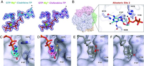
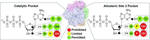
SAMHD1 is a deoxynucleoside triphosphate triphosphohydrolase (dNTPase) that depletes cellular dNTPs in noncycling cells to promote genome stability and to inhibit retroviral and herpes viral replication. In addition to being substrates, cellular nucleotides also allosterically regulate SAMHD1 activity. Recently, it was shown that high expression levels of SAMHD1 are also correlated with significantly worse patient responses to nucleotide analog drugs important for treating a variety of cancers, including acute myeloid leukemia (AML). In this study, we used biochemical, structural, and cellular methods to examine the interactions of various cancer drugs with SAMHD1. We found that both the catalytic and the allosteric sites of SAMHD1 are sensitive to sugar modifications of the nucleotide analogs, with the allosteric site being significantly more restrictive. We crystallized cladribine-TP, clofarabine-TP, fludarabine-TP, vidarabine-TP, cytarabine-TP, and gemcitabine-TP in the catalytic pocket of SAMHD1. We found that all of these drugs are substrates of SAMHD1 and that the efficacy of most of these drugs is affected by SAMHD1 activity. Of the nucleotide analogs tested, only cladribine-TP with a deoxyribose sugar efficiently induced the catalytically active SAMHD1 tetramer. Together, these results establish a detailed framework for understanding the substrate specificity and allosteric activation of SAMHD1 with regard to nucleotide analogs, which can be used to improve current cancer and antiviral therapies.

Keywords: SAMHD1; allosteric regulation; dNTPase; nucleotide analog drugs; substrate selection.


Conflict of interest statement

The authors declare no conflict of interest.

Figures

Fig. 1.
Fig. 1.
Substrate specificity of SAMHD1 is determined by 2′ sugar moiety. (A) Chemical structures of nucleoside analogs used in this study. (B) 2Fo-Fc electron density (σ = 1.0) for the nucleotide analog drugs crystallized in the catalytic pocket of SAMHD1. Black asterisks indicate sites of modifications. (C, Top) Transparent surface view of SAMHD1 tetramer with each subunit in a different color. Selected allosteric nucleotides are shown in red sticks, and a nucleotide in a catalytic pocket is shown in blue sticks. (C, Bottom) Superposition of all of the nucleotide analogs bound to the SAMHD1 catalytic pocket. SAMHD1 backbone is shown as coils with side chains shown as sticks. Cladribine-TP (cyan), clofarabine-TP (magenta), fludarabine-TP (green), vidarabine-TP (wheat), cytarabine-TP (purple), and gemcitabine-TP (orange) are shown as sticks. (D) Water networks (shown as red spheres) observed for each nucleotide analog bound to the SAMHD1 catalytic site. Black dashed lines indicate hydrogen bonds.
Fig. 2.
Fig. 2.
Gemcitabine-TP but not [(2′R)-2′-F]-dCTP is hydrolyzed by SAMHD1 in vitro. (A) [(2′S)-2′-OH)] of cytarabine-TP is stabilized by residues Y374 and Y315 through van der Waals interactions. Transparent surface of SAMHD1 is shown with key residues in sticks. (B) The 2′,2′-difluorine sugar modification of gemcitabine-TP is stabilized by van der Waals interactions with residues Y374 and Y315 to compensate potential close contact (yellow caution triangle) between the [(2′R)-2′-F] atom and residue L150 in the catalytic site. (C) HPLC-based activity assay measuring product produced by preassembled SAMHD1 tetramers in the presence of 25 to 1,600 μM nucleotide analog substrates. Error bars represent SEM of three independent experiments. (D, Left) dNTPase activity of SAMHD1 over the course of 15 min was measured using a malachite green assay. Product is normalized to SAMHD1 concentration (nmol PO4/nmol SAMHD1). SAMHD1 tetramers were preassembled with 250 μM GTP and dATP and then diluted 100-fold into 125 μM gemcitabine-TP, cytarabine-TP, dCTP, CTP, [(2′S)-2′-F]-dCTP, [(2′R)-2′-F]-dCTP, or buffer. Error bars represent SEM of three independent experiments. (D, Right) Chemical structures of [(2′S)-2′-F]-dCTP and [(2′R)-2′-F]-dCTP analogs. (E) A rigid body model of [(2′R)-2′-F]-dCTP (gray sticks) in the catalytic pocket potentially creates a clash (red cross) with residue L150.
Fig. 3.
Fig. 3.
SAMHD1 depletes several TPs of nucleoside analogs in vivo. (A) Correlations of cytarabine, clofarabine, fludarabine, cladribine, and gemcitabine concentrations inhibiting 50% of cell viability (IC50) and relative protein expression levels of SAMHD1 in 13 AML cell lines. Relative expression levels (ratios of a SAMHD1/β-actin) are shown as arbitrary units (a.u.). Ratio of SAMHD1/β-actin for parental THP-1 cells is set to 1, and ratios of other cell lines are set relative to it. Filled circles represent mean values, and error bars indicate SD of three independent experiments. Correlations were analyzed using linear or log-linear regression models. (B) Cytarabine, fludarabine, clofarabine, cladribine, or gemcitabine IC50 values of THP-1 KO, THP-1 Control cells, or parental THP-1 cells exposed to VSV-G pseudotyped VLPs carrying either lentiviral Vpr (Vpr-VLP, control) or SAMHD1-degrading Vpx proteins (Vpx-VLP). The bars represent mean values, and the error bars are SD of three independent experiments. The numbers above indicate factor of decrease of the IC50 values in the absence of SAMHD1. Statistical analyses were performed using unpaired two-tailed Student’s t test comparing treated samples with untreated control. n.s., no statistical significance. ***P < 0.001. (C) Representative liquid chromatography tandem mass spectrometry (LC-MS/MS) measurements of cytarabine-TP, fludarabine-TP, clofarabine-TP, cladribine-TP, or gemcitabine-TP in THP-1 KO cells (black) and THP-1 control cells (red).
Fig. 4.
Fig. 4.
The allosteric sites of SAMHD1 are highly restrictive. (A) Size-exclusion chromatography elution profile of SAMHD1 in the presence of 0.5 mM GTP and 4 mM of color-coded nucleotide analog. (B) SV-AUC analysis of SAMHD1 in the absence of nucleotides or the presence of dGTP, GTP with clofarabine-TP, or GTP with cladribine-TP at a final concentration of 150 μM. (C) Malachite green activity assay performed in the presence of 125 μM GTP and 125 μM dCTP, dATP, nucleotide analog, or buffer. Error bars represent SEM of three independent experiments. (D) Malachite green activity assay measuring the hydrolysis of dATP by SAMHD1 tetramers preassembled in the presence of 125 μM GTP and 6.3 to 3,200 μM dATP, cladribine-TP, clofarabine-TP, or ATP. Error bars represent SEM of three independent experiments.
Fig. 5.
Fig. 5.
Structures of cladribine-TP and clofarabine-TP bound to Allo-site 2 of SAMHD1. (A) 2Fo-Fc electron density (σ = 1.0) for GTP, cladribine-TP, and clofarabine-TP in the allosteric pocket of SAMHD1. Black asterisks indicate sites of modifications, and black dotted lines indicate hydrogen bonds. (B, Left) Transparent surface view of the SAMHD1 tetramer. (B, Right) Overlay of cladribine-TP (cyan) and clofarabine-TP (pink) in Allo-site 2. The main chain of SAMHD1 is shown as tubes, with selected residues and nucleotides represented as sticks. Residues important for gating the 2′-atom are highlighted in thicker sticks. Black asterisks indicate sites of modification. (C) The structure of cladribine-TP (cyan, sticks) in Allo-site 2 with V156, F157, and H376 shown as sticks under semitransparent surface of SAMHD1. (D) The structure of clofarabine-TP (pink, sticks) in Allo-site 2 with close contacts between the [(2′S)-2′-F] atom and V156, F157, and H376 highlighted with caution triangles and yellow dashed lines. (E) A rigid body model of cytarabine-TP in Allo-site 2 with potential steric clashes between the [(2′S)-2′-OH] group and V156 and H376 highlighted with a red cross and red dashed lines. (F) A rigid body model of gemcitabine-TP in Allo-site 2 with a potential steric clash between the [(2′R)-2′-F] atom and F157 highlighted with a red cross and red dashed lines.
Fig. 6.
Fig. 6.
Summary of the effects of the 2′ sugar moiety on nucleotide analog binding to the catalytic and allosteric sites of SAMHD1. (Middle) Transparent surface view of SAMHD1 tetramer. (Left) In the catalytic pocket of SAMHD1, while small substitutions such as fluorine atoms at the 2′R and 2′S positions of the sugar are permitted (green circle), their access to the 2′R position is limited (yellow circle). Larger modifications, such as hydroxyl groups, are permitted in the 2′S position, but not the 2′R position (red circle). (Right) In Allo-site 2 of SAMHD1, hydroxyl groups are prohibited in both 2′R and 2′S positions of the sugar moiety. Fluorine atoms have limited access to the 2′S position, but they are prohibited from the 2′R position. Small base modifications, such as fluorine or chlorine atoms, are tolerated in both Allo-site 2 and the catalytic site.

References

    1. Franzolin E, et al. The deoxynucleotide triphosphohydrolase SAMHD1 is a major regulator of DNA precursor pools in mammalian cells. Proc Natl Acad Sci USA. 2013;110:14272–14277. - PMC - PubMed
    1. Goldstone DC, et al. HIV-1 restriction factor SAMHD1 is a deoxynucleoside triphosphate triphosphohydrolase. Nature. 2011;480:379–382. - PubMed
    1. Lahouassa H, et al. SAMHD1 restricts the replication of human immunodeficiency virus type 1 by depleting the intracellular pool of deoxynucleoside triphosphates. Nat Immunol. 2012;13:223–228. - PMC - PubMed
    1. Descours B, et al. SAMHD1 restricts HIV-1 reverse transcription in quiescent CD4(+) T-cells. Retrovirology. 2012;9:87. - PMC - PubMed
    1. Bermejo M, et al. Dasatinib inhibits HIV-1 replication through the interference of SAMHD1 phosphorylation in CD4+ T cells. Biochem Pharmacol. 2016;106:30–45. - PubMed

Publication types

MeSH terms

Substances