

Staggered Hox expression is more widespread among molluscs than previously appreciated
- PMID: 30305436
- PMCID: PMC6191704
- DOI: 10.1098/rspb.2018.1513
Staggered Hox expression is more widespread among molluscs than previously appreciated
Abstract
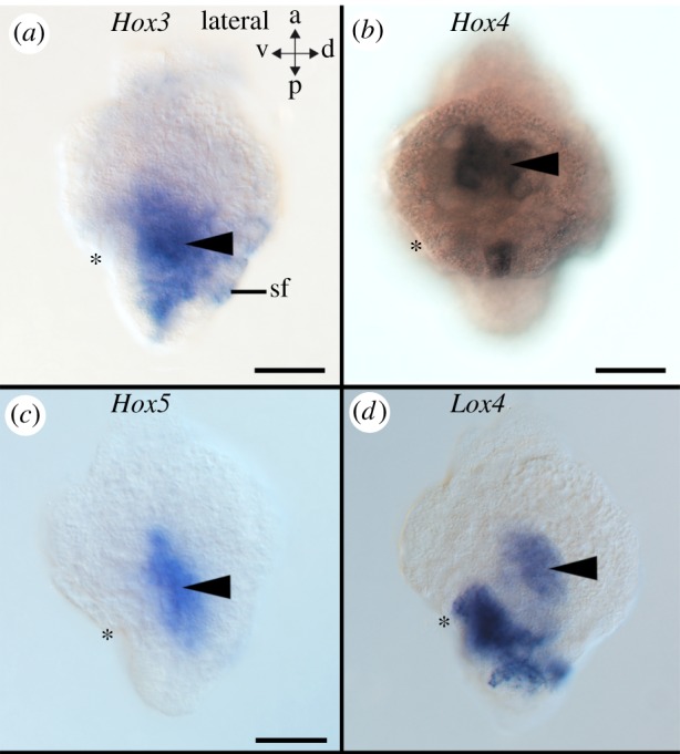
Hox genes are expressed along the anterior-posterior body axis in a colinear fashion in the majority of bilaterians. Contrary to polyplacophorans, a group of aculiferan molluscs with conserved ancestral molluscan features, gastropods and cephalopods deviate from this pattern by expressing Hox genes in distinct morphological structures and not in a staggered fashion. Among conchiferans, scaphopods exhibit many similarities with gastropods, cephalopods and bivalves, however, the molecular developmental underpinnings of these similar traits remain unknown. We investigated Hox gene expression in developmental stages of the scaphopod Antalis entalis to elucidate whether these genes are involved in patterning morphological traits shared by their kin conchiferans. Scaphopod Hox genes are predominantly expressed in the foot and mantle but also in the central nervous system. Surprisingly, the scaphopod mid-stage trochophore exhibits a near-to staggered expression of all nine Hox genes identified. Temporal colinearity was not found and early-stage and late-stage trochophores, as well as postmetamorphic individuals, do not show any apparent traces of staggered expression. In these stages, Hox genes are expressed in distinct morphological structures such as the cerebral and pedal ganglia and in the shell field of early-stage trochophores. Interestingly, a re-evaluation of previously published data on early-stage cephalopod embryos and of the gastropod pre-torsional veliger shows that these developmental stages exhibit traces of staggered Hox expression. Considering our results and all gene expression and genomic data available for molluscs as well as other bilaterians, we suggest a last common molluscan ancestor with colinear Hox expression in predominantly ectodermal tissues along the anterior-posterior axis. Subsequently, certain Hox genes have been co-opted into the patterning process of distinct structures (apical organ or prototroch) in conchiferans.
Keywords: Lophotrochozoa; Polyplacophora; cephalopod; gastropod; gene expression; nervous system.
© 2018 The Authors.
Conflict of interest statement
We declare we have no competing interests.
Figures


Similar articles
-
Non-collinear Hox gene expression in bivalves and the evolution of morphological novelties in mollusks.Sci Rep. 2021 Feb 11;11(1):3575. doi: 10.1038/s41598-021-82122-6. Sci Rep. 2021. PMID: 33574385 Free PMC article.
-
Unexpected co-linearity of Hox gene expression in an aculiferan mollusk.BMC Evol Biol. 2015 Aug 5;15:151. doi: 10.1186/s12862-015-0414-1. BMC Evol Biol. 2015. PMID: 26243538 Free PMC article.
-
The ParaHox gene Gsx patterns the apical organ and central nervous system but not the foregut in scaphopod and cephalopod mollusks.Evodevo. 2015 Dec 29;6:41. doi: 10.1186/s13227-015-0037-z. eCollection 2015. Evodevo. 2015. PMID: 26715985 Free PMC article.
-
Hox and ParaHox genes: a review on molluscs.Genesis. 2014 Dec;52(12):935-45. doi: 10.1002/dvg.22839. Epub 2014 Nov 28. Genesis. 2014. PMID: 25394269 Review.
-
Hox cluster genes and collinearities throughout the tree of animal life.Int J Dev Biol. 2018;62(11-12):673-683. doi: 10.1387/ijdb.180162sg. Int J Dev Biol. 2018. PMID: 30604837 Review.
Cited by
-
Gene Recruitments and Dismissals in the Argonaut Genome Provide Insights into Pelagic Lifestyle Adaptation and Shell-like Eggcase Reacquisition.Genome Biol Evol. 2022 Nov 4;14(11):evac140. doi: 10.1093/gbe/evac140. Genome Biol Evol. 2022. PMID: 36283693 Free PMC article.
-
Non-collinear Hox gene expression in bivalves and the evolution of morphological novelties in mollusks.Sci Rep. 2021 Feb 11;11(1):3575. doi: 10.1038/s41598-021-82122-6. Sci Rep. 2021. PMID: 33574385 Free PMC article.
-
Octopod Hox genes and cephalopod plesiomorphies.Sci Rep. 2023 Sep 19;13(1):15492. doi: 10.1038/s41598-023-42435-0. Sci Rep. 2023. PMID: 37726311 Free PMC article.
-
The genome of the venomous snail Lautoconus ventricosus sheds light on the origin of conotoxin diversity.Gigascience. 2021 May 25;10(5):giab037. doi: 10.1093/gigascience/giab037. Gigascience. 2021. PMID: 34037232 Free PMC article.
-
Signatures of Divergence, Invasiveness, and Terrestrialization Revealed by Four Apple Snail Genomes.Mol Biol Evol. 2019 Jul 1;36(7):1507-1520. doi: 10.1093/molbev/msz084. Mol Biol Evol. 2019. PMID: 30980073 Free PMC article.
References
-
- Vinther J. 2015. The origins of molluscs. Palaeontology 58, 19–34. (10.1111/pala.12140) - DOI
-
- Parkhaev PY. 2017. Origin and the early evolution of the phylum Mollusca. Paeont. J. 51, 663–686.
-
- Wanninger A, Wollesen T. 2015. Mollusca in evolutionary developmental biology of invertebrates, vol. 2 lophotrochozoa (ed. Wanninger A.), pp. 103–153. Berlin, Germany: Springer.
Publication types
MeSH terms
Associated data
Grants and funding
LinkOut - more resources
Full Text Sources
