. 2018 Oct 10;9(10):1039.
doi: 10.1038/s41419-018-1099-3.

Diosgenin promotes antitumor immunity and PD-1 antibody efficacy against melanoma by regulating intestinal microbiota

Affiliations

Diosgenin promotes antitumor immunity and PD-1 antibody efficacy against melanoma by regulating intestinal microbiota

Mengxue Dong et al. Cell Death Dis. .

Abstract

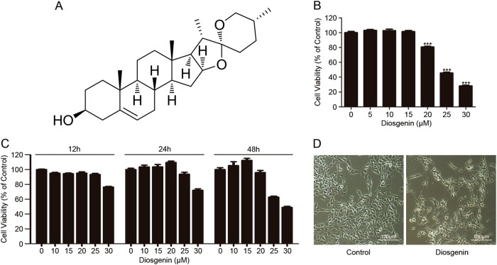
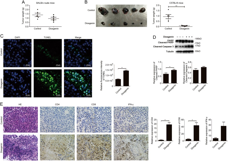
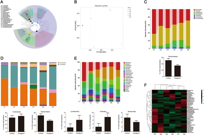
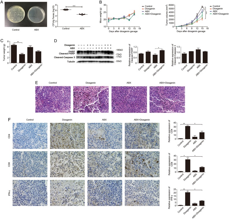
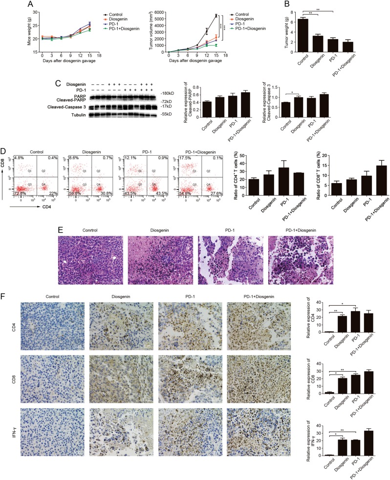
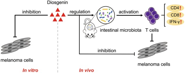
Diosgenin, a natural steroidal saponin, can exert antitumor effect by regulating immune function and improving intestinal microbiota. The response to anti-PD-1 immunotherapy is associated with intestinal microbiota and effector T cells in tumor microenvironment. We hypothesize that the modulation of diosgenin on intestinal microbiota can facilitate antitumor immunity and the therapeutic efficacy of PD-1 antibody. In melanoma-bearing C57BL/6 mice, we observed that the anti-melanoma effect of diosgenin relied more on antitumor immunity than direct tumor inhibition activity evidenced by obvious CD4+/CD8+ T-cell infiltration and IFN-γ expression in tumor tissues, and it could improve the compositions of intestinal microbiota. Antibiotics impaired the therapeutic efficacy and immunity responses of diosgenin through disturbing intestinal microbiota, indicating the importance of intestinal microbiota in diosgenin's in vivo antitumor activity. More importantly, the combined administration of PD-1 antibody with diosgenin aggravated the tumor necrosis and apoptosis by eliciting augmented T-cell responses. Taken together, diosgenin can be used as a microecological regulator to induce antitumor immunity and improve the efficacy of immune checkpoint antibody, making it more suitable for the treatment of malignant tumors.


Conflict of interest statement

The authors declare that they have no conflict of interest.

Figures

Fig. 1
Fig. 1. Diosgenin induced cytotoxicity in B16F10 cells.
a The structure of diosgenin. b B16F10 cells were treated with various concentrations of diosgenin for 48 h, and then MTT assay was applied to measure the cell viability (IC50 of diosgenin = 25 μM). All these data were represented as mean ± SD (***P < 0.001). c B16F10 cells were exposed to different concentrations of diosgenin for indicated times. Cell viability was measured by MTT assay. d B16F10 cells were administered with 25 μM diosgenin for 48 h and the morphological alterations of cells were observed by an inverted microscope
Fig. 2
Fig. 2. In vivo anti-melanoma activity of diosgenin dependent on its antitumor immunity.
The mice were subcutaneously injected with 2×10⁵ of B16F10 cells. a The tumor weight of nude mice daily treated with normal saline (control) and diosgenin (20 mg/kg) respectively for 2 weeks. The results were represented as means ± SD. be The tumor-bearing C57BL/6 mice were treated with or without diosgenin (20 mg/kg) everyday. After administration for 2 weeks, the tumors were excised (b, left), and the tumor weight was shown in (b, right). c Apoptosis of tumor tissues from C57BL/6 mice was assayed with TUNEL and observed under fluorescence microscope. The quantitation analysis of apoptotic tumor cells was performed by ImageJ software and presented in bar charts. d The expression levels of PARP, cleaved-PARP, and cleaved-caspase 3 in tumor tissues from C57BL/6 mice were checked by western blot analysis. Densitometric values were quantified by ImageJ software. e Representative H&E staining images of tumor tissues were shown in the left panel, and the expressions of CD4, CD8, and IFN-γ in tumor tissues were detected by immunohistochemistry. The quantitation analysis was performed by ImageJ software. All these data were presented as mean ± SD (*P < 0.05, **P < 0.01)
Fig. 3
Fig. 3. Diosgenin had a regulatory effect on intestinal microbiota in C57BL/6 mice.
The C57BL/6 mice were administered by gavage with normal saline (control) or diosgenin (20 mg/kg) everyday; the feces of mice were collected after 2 weeks to detect the intestinal microbiota. C1, C2, and C3 belonged to the control group, D1, D2, and D3 belonged to the diosgenin-treated group. a The classification tree of sample population based on GraPhlAn. b Two-dimension ordination graph of PCA analysis, in which distances between the samples represented the differences of them. ce The community taxonomic composition and abundance distribution map from phylum to genus. The taxonomic units were distinguished by different colors, and the length of the column represented the relative abundance of each unit. c At the phylum level, the abundance of Bacteroidetes was counted. d At the order level, the abundances of Clostridiales and Bacteroidales were analyzed. e According to the level of genus, the abundances of Lactobacillus, Sutterella, and Bacteroides were respectively analyzed. All the results were presented as mean ± SD. f The heatmap with log10-transformation of relative abundance at the genus level in the intestine that was indicated from green (less) to red (more)
Fig. 4
Fig. 4. Diosgenin triggered anti-melanoma immune effect by modulating intestinal microbiota in tumor-bearing C57BL/6 mice.
The C57BL/6 mice received antibiotic cocktail (ABX) 2 weeks before tumor inoculation. After subcutaneously injected with 1×10⁶ of B16F10 cells, the mice were daily treated with or without diosgenin (20 mg/kg). a The feces of mice were cultivated on LB solid plates (left) post-ABX administration, and the number of bacterial colonies was shown in the right panel. b The mice weight and the tumor volume of four groups were detected throughout the experiment. After mice were sacrificed, the tumor weight was measured (c). d The expression levels of PARP, cleaved-PARP, and cleaved-caspase 3 in tumor tissues were assessed by western blot analysis. Densitometric values were quantified by ImageJ software. e Representative H&E staining images of tumor tissues from each group. f Immunohistochemical analysis of tumor tissues, and the expression levels of CD4, CD8, and IFN-γ were observed under microscope. The quantitation analysis was performed by ImageJ software and presented in bar charts. All these data were represented as mean ± SD (*P < 0.05, **P < 0.01, ***P < 0.001)
Fig. 5
Fig. 5
Combined administration of diosgenin and PD-1 mAb achieved the best antitumor effect. After subcutaneously injected with 1×10⁶ of B16F10 cells, the C57BL/6 mice were daily treated with or without diosgenin (20 mg/kg), and then injected intraperitoneally with PD-1 mAb on days 6, 9, and 12 post-tumor implantation. a The mice weight and the tumor volume of four groups were detected throughout the experiment. b After the whole experiment, the tumor weight was measured. c The expression levels of PARP, cleaved-PARP, and cleaved-caspase 3 in tumor tissues were assessed by western blot analysis. Densitometric values were quantified by ImageJ software. d The number of CD4+/CD8+ T cells in tumor tissues was analyzed by flow cytometry, and the percentage of these cells was presented in bar charts as mean ± SD. e Representative H&E staining images of tumor tissues from each group. f The expression levels of CD4, CD8, and IFN-γ in tumor tissues were determined by immunohistochemistry, and the quantitation analysis was performed by ImageJ software. All these data were presented as mean ± SD (*P < 0.05, **P < 0.01, ***P < 0.001). PD-1 indicates PD-1 monoclonal antibody
Fig. 6
Fig. 6. Overview of anti-melanoma effect induced by diosgenin in vitro and in vivo.
Diosgenin can inhibit the growth of B16F10 melanoma cells in vitro. In vivo, the anti-melanoma effect of diosgenin is mainly dependent on antitumor immunity which is induced by its regulation of intestinal microbiota in C57BL/6 mice

References

    1. Wolchok JD, Chan TA. Cancer: antitumour immunity gets a boost. Nature. 2014;515:496–498. doi: 10.1038/515496a. - DOI - PMC - PubMed
    1. Sharma P, Allison JP. The future of immune checkpoint therapy. Science. 2015;348:56–61. doi: 10.1126/science.aaa8172. - DOI - PubMed
    1. Weiner GJ. Building better monoclonal antibody-based therapeutics. Nat. Rev. Cancer. 2015;15:361–370. doi: 10.1038/nrc3930. - DOI - PMC - PubMed
    1. Chen L, Han X. Anti-PD-1/PD-L1 therapy of human cancer: past, present, and future. J. Clin. Invest. 2015;125:3384–3391. doi: 10.1172/JCI80011. - DOI - PMC - PubMed
    1. Robert C, et al. Pembrolizumab versus ipilimumab in advanced melanoma. N. Engl. J. Med. 2015;372:2521–2532. doi: 10.1056/NEJMoa1503093. - DOI - PubMed

Publication types

MeSH terms