Review
. 2018 Sep;10(Suppl 24):S2837-S2847.
doi: 10.21037/jtd.2018.02.18.

Anatomy of the atrial septum and interatrial communications

Affiliations
Review

Anatomy of the atrial septum and interatrial communications

Nitha Naqvi et al. J Thorac Dis. 2018 Sep.

Abstract

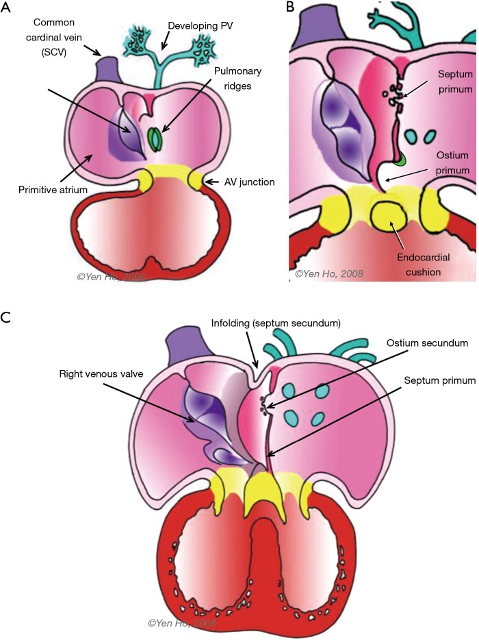
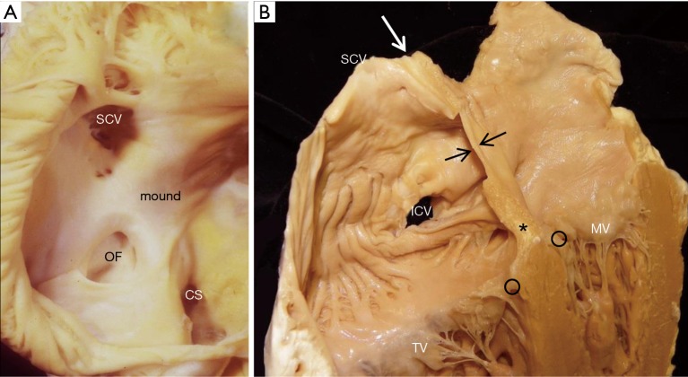
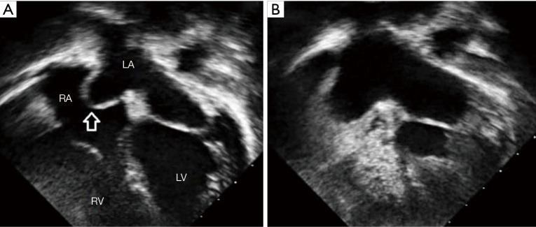
Deficiencies in the septum separating the two atrial chambers are among the most common of congenital heart malformations. This article reviews the developmental aspects of the partitioning of the primitive atrium into right and left atrial chambers, the anatomical components of the atrial septum, and deficiencies that produce the various types of interatrial communications. Knowledge of the components of the true atrial septum in the developed heart clarifies the morphology of various types of interatrial communications. The oval fossa defect (also termed secundum ASD) is located within the true septum. The patent foramen ovale (PFO) is a tunnel-like passageway between the free edge of the overlapping ovale fossa valve and its muscular rim. Other defects such as superior and inferior sinus venosus defects, coronary sinus defects, and ostium primum defects lie outside the area of the true septum.

Keywords: Atrial septal defect (ASD); coronary sinus defect; patent foramen ovale (PFO); sinus venosus defect.


Conflict of interest statement

Conflicts of Interest: The authors have no conflicts of interest to declare.

Figures

Figure 1
Figure 1
Septation of the common atrial chamber. (A) The primitive atrial chamber opens to the developing ventricles through the atrioventricular (AV) canal. The right and left venous valves mark the border between the sinus venosus and the primitive atrium. The developing pulmonary veins enters the atrium at the pulmonary pit; (B) as the septum primum extends toward the atrioventricular canal, its upper portion breaks down to form the ostium secundum; (C) immediately to the right of the septum primum, the atrial wall folds to form the septum secundum. The inferior ends of the right and left venous valve fuse with tissues of the septum primum and spina vestibuli (Figure adapted from Ho SY. Embryology and anatomy of the atrial septum. In: Transseptal Catheterization and Interventions. Thakur R, Natale A, editors; Cardiotext 2010, Minnesota USA. Pages 11–26).
Figure 2
Figure 2
The component parts of the definitive atrial septum (Figure adapted from Ho SY. Embryology and anatomy of the atrial septum. In: Transseptal Catheterization and Interventions. Thakur R, Natale A, editors; Cardiotext 2010, Minnesota USA. Pages 11–26).
Figure 3
Figure 3
Profile of the septal plane in situ. (A) A heart sectioned in simulated transoesophageal plane showing the plane of the atrial septum (asterisks); (B) Transesophageal echocardiogram showing the oblique plane of the atria septum. AV, aortic valve; DAo, descending aorta; LA, left atrium; Oes, oesophagus; RA, right atrium; RVOT, right ventricular outflow tract.
Figure 4
Figure 4
The true septum. (A) A heart specimen with the right atrium opened to display the atrial septal surface enface. The true septum is limited to the area of the oval fossa (OF) and the immediate margin of the surrounding muscular rim; (B) A heart specimen cut in four-chamber plane shows the configuration of the atrial septum which is the thin area (in between arrows) bordered by the thicker muscular rim. This cut through the infolded atrial wall shows epicardial fat filling the fold (white arrow). The septal insertions of the mitral valve (MV) and tricuspid valve (TV) hinge lines (o) are offset and in between, where atrial septal structures lie above the ventricular septum, there is an area of fatty tissues from the epicardium (asterisk). CS, coronary sinus; ICV, inferior caval vein; OF, oval fossa; SCV, superior caval vein.
Figure 5
Figure 5
Location of patent foramen ovale (PFO). (A) Right atrial view of the atrial septum enface with a probe passing from the right atrium through the PFO into the left atrium. Note the location of the PFO at the antero-cephalad margin of the fossa; (B) left atrial view showing the probe directed toward the anterior wall of the left atrium (LA), immediately behind the aorta (Ao). There is a dimple where the atrial wall is thin (arrow); (C) echocardiogram of subcostal view of the atria illustrating a patent foramen ovale (red arrow). CS, coronary sinus; LV, left ventricle; RAA, right atrial appendage; RV, right ventricle.
Figure 6
Figure 6
Septal aneurysm. (A) Echocardiogram of a 4 chamber view illustrating an interatrial septum (shown by the arrow) bowing into the right atrium; (B) contrast echocardiogram of the same patient demonstrating that the interatrial septum is intact and there is absence of a PFO shunt. RA, right atrium; LA, left atrium; RV, right ventricle; LV, left ventricle; PFO, patent foramen ovale.
Figure 7
Figure 7
Types of interatrial communications. (A) The most common form of interatrial communication is at the site of the oval fossa making it a true atrial septal defect (ASD). The superior (Sup) and inferior (Inf) sinus venosus (SV) defects, coronary sinus (cs) defects, and atrioventricular septal defects (AVSD) are outside the atrial septum although they permit interatrial shunting; (B) an enface view of a fenestrated oval fossa defect. CS, coronary sinus; ICV, inferior caval vein; SCV, superior caval vein; TV, tricuspid valve.
Figure 8
Figure 8
Superior sinus venosus defect. (A) Diagram showing the location of the superior sinus venosus defect (double arrows) above the atrial septum. The oval fossa is intact and its superior rim encloses epicardial fat (blue). The right superior pulmonary vein (RSPV) connects anomalously to the entrance of the superior caval vein (SCV) into the right atrium; (B) a heart specimen with the parietal right atrial wall removed to show the superior sinus venosus defect (blue star) beneath the entrance of the superior caval vein. The true atrial septum, the oval fossa(OF), is intact; (C) echocardiogram of a subcostal bicaval view illustrating a superior sinus venosus defect (red arrow) with the superior caval vein overriding the interatrial septum. CS, coronary sinus; ICV, inferior caval vein; LA, left atrium; OF, oval fossa; RA, right atrium.

Similar articles

Cited by

References

    1. van der Linde D, Konings EE, Slager MA, et al. Birth prevalence of congenital heart disease worldwide. A systematic review and meta-analysis. J Am Coll Cardiol 2011;58:2241-7. - PubMed
    1. Gittenberger-de Groot A, Bartelings MM, Deruitter MC, et al. Basics of cardiac development for understanding of congenital heart malformations. Pediatr Res 2005;57:169-76. 10.1203/01.PDR.0000148710.69159.61 - DOI - PubMed
    1. Kim JS, Virágh S, Moorman AF, et al. Development of the myocardium of the atrioventricular canal and the vestibular spine in the human heart. Circ Res 2001;88:395-402. 10.1161/01.RES.88.4.395 - DOI - PubMed
    1. Lamers WH, Moorman AF. Cardiac septation. A late contribution of the embryonic primary myocardium to heart morphogenesis. Circ Res 2002;91:93-103. 10.1161/01.RES.0000027135.63141.89 - DOI - PubMed
    1. Mommersteeg MT, Soufan AT, de Lange FJ, et al. Two distinct pools of mesenchyme contribute to the development of the atrial septum. Circ Res 2006;99:351-3. 10.1161/01.RES.0000238360.33284.a0 - DOI - PubMed