Review
. 2018 Sep;10(Suppl 24):S2874-S2881.
doi: 10.21037/jtd.2018.08.111.

Atrial septal defect closure: indications and contra-indications

Affiliations
Review

Atrial septal defect closure: indications and contra-indications

Alain Fraisse et al. J Thorac Dis. 2018 Sep.

Abstract

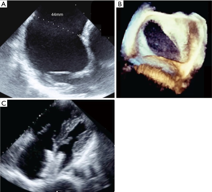
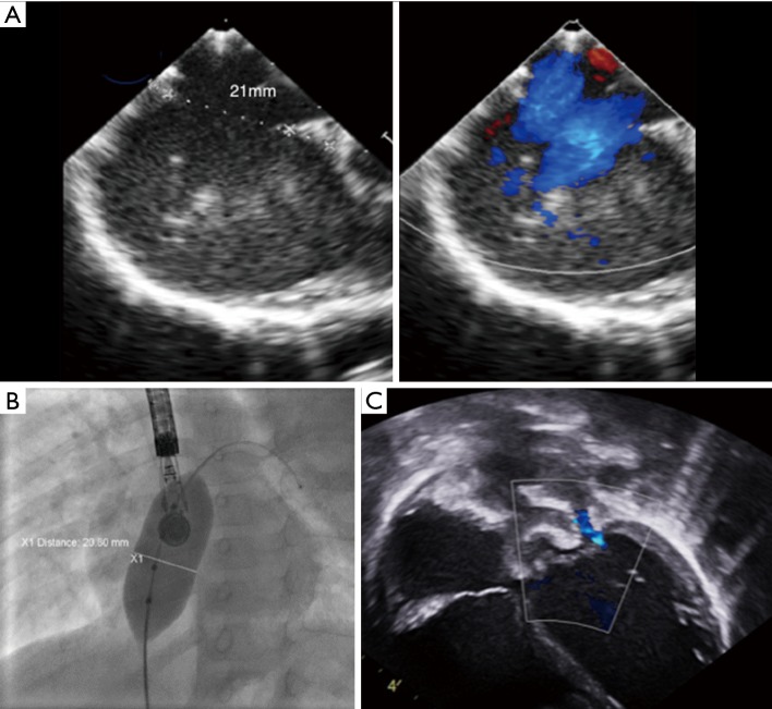
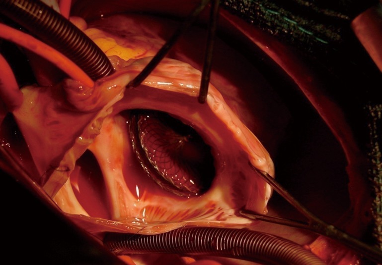
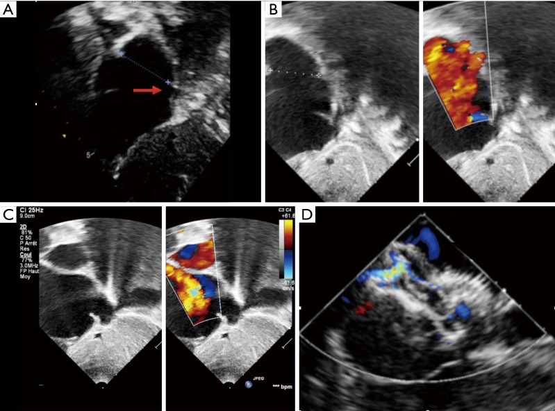
Transcatheter closure has become an accepted alternative to surgical repair for ostium secundum atrial septal defects (ASD). However, large ASDs (>38 mm) and defects with deficient rims are usually not offered transcatheter closure but are referred for surgical closure. Transcatheter closure also remains controversial for other complicated ASDs with comorbidities, additional cardiac features and in small children. This article not only provides a comprehensive, up-to-date description of the current indications and contra-indications for ASD device closure, but also further explores the current limits for transcatheter closure in controversial cases. With the devices and technology currently available, several cohort studies have reported successful percutaneous closure in the above-mentioned complex cases. However the feasibility and safety of transcatheter technique needs to be confirmed through larger studies and longer follow-up.

Keywords: Atrial septal defect (ASD); cardiac catheterisation; closure.


Conflict of interest statement

Conflicts of Interest: Alain Fraisse is a consultant and proctor for Abbott Inc. and Occlutech Inc. The other authors have no conflicts of interest to declare.

Figures

Figure 1
Figure 1
Transcatheter closure of a very large oval atrial septal defect. (A) Very large atrial septal defect with maximum diameter measured at 44 mm on transoesophageal echocardiography; (B) 3D reconstruction demonstrates the crescent shape of the defect with a much smaller transverse diameter; (C) transthoracic echocardiography in 4-chambers view shows good position of the device after successful closure with a 40 mm Amplatzer ASO. ASO, Amplatzer septal occluder.
Figure 2
Figure 2
Large atrial septal defect that is measured at 21 mm on transesophageal echocardiography (A) in a 9.7 kg, 11-month-old infant with failure to thrive. (B) The transoesophageal echocardiography diameter is confirmed with the sizing balloon under fluoroscopy. After a 22 mm Amplatzer ASO is successfully implanted post-procedure transthoracic echocardiography in 4 chambers view confirms good position of the device and demonstrates a new mild mitral regurgitation that is functional, due to the sudden increase in left ventricular volume (C). This mild mitral regurgitation will disappear after a few weeks on follow-up echocardiography.
Figure 3
Figure 3
Intra-operative picture after device embolization in a 9-year-old child with moderate atrial septal defect showing the complete absence of tissue (arrow) between the inferior vena cava (where the inferior vena cava is cannulated) and the defect. The device was initially well-positioned but embolization probably occurred after the device slipped toward the inferior vena cava because of the absence of any rim.
Figure 4
Figure 4
Difference on echocardiography between postero inferior and posterior rim deficiency. (A) Subcostal view with transthoracic echocardiography shows very large atrial septal defect with complete posteroinferior rim deficiency toward the inferior vena cava (arrow). This child will be referred for surgery. (B) A subcostal view in another 14 kg child shows a very large defect with complete posterior rim deficiency. However subsequent subcostal view toward the inferior vena cava in (C) demonstrate a posteroinferior rim (arrow). This will successfully be closed with good position of the device under transoesophageal echocardiography (D).
Figure 5
Figure 5
Transcatheter closure of an “octopus defect” mimicking multiple defects. (A) Transoesophageal echocardiography view showing multiple secundum atrial septal defects; (B) transthoracic echocardiography demonstrates successful closure of the defect with a single large device and no residual shunt.
Figure 6
Figure 6
Transcatheter closure of multiple atrial septal defect with 2 devices. (A) 3D reconstruction with transoesophageal echocardiography showing a residual defect after a 32 mm Amplatzer ASO has been positioned through the biggest atrial septal defect; (B) successful closure after implantation of a 30 mm Amplatzer cribriform device overlapping the original 32 mm ASO. ASO, Amplatzer septal occluder.
Figure 7
Figure 7
Fluoroscopy in left anterior oblique view showing closure of a large atrial septal defect in a patient with dextrocardia and situs inversus.
Figure 8
Figure 8
Fluoroscopy in left anterior oblique view showing closure of an atrial septal defect though transhepatic access.

References

    1. Mills NL, King TD. Nonoperative closure of left-to-right shunts. J Thorac Cardiovasc Surg 1976;72:371-8. - PubMed
    1. Du ZD, Hijazi ZM, Kleinman CS, et al. Comparison between transcatheter and surgical closure of secundum atrial septal defect in children and adults: results of a multicenter nonrandomized trial. J Am Coll Cardiol 2002;39:1836-44. 10.1016/S0735-1097(02)01862-4 - DOI - PubMed
    1. Butera G, Romagnoli E, Carminati M, et al. Treatment of isolated secundum atrial septal defects: impact of age and defect morphology in 1,013 consecutive patients. Am Heart J 2008;156:706-12. 10.1016/j.ahj.2008.06.008 - DOI - PubMed
    1. Warnes CA, Williams RG, Bashore TM, et al. ACC/AHA 2008 guidelines for the management of adults with congenital heart disease: a report of the American College of Cardiology/American Heart Association Task Force on Practice Guidelines (Writing Committee to Develop Guidelines on the Management of Adults With Congenital Heart Disease). Developed in Collaboration With the American Society of Echocardiography, Heart Rhythm Society, International Society for Adult Congenital Heart Disease, Society for Cardiovascular Angiography and Interventions, and Society of Thoracic Surgeons. J Am Coll Cardiol 2008;52:e143-e263. 10.1016/j.jacc.2008.10.001 - DOI - PubMed
    1. Baruteau AE, Hascoët S, Fraisse A. Transthoracic echocardiography is a safe alternative for assessment and guidance of transcatheter closure of secundum atrial septal defect in children. J Thorac Dis 2017;9:1247-56. 10.21037/jtd.2017.04.47 - DOI - PMC - PubMed