

Review
doi: 10.21037/jtd.2018.07.116.
Surgical closure of atrial septal defects
Affiliations
- PMID: 30305953
- PMCID: PMC6174143
- DOI: 10.21037/jtd.2018.07.116
Item in Clipboard
Review
Surgical closure of atrial septal defects
J Thorac Dis.
2018 Sep.
Abstract
Surgical repair of an atrial septal defect (ASD) is a safe and effective operation with little to no morbidity and mortality. In an effort to reduce the trauma of surgery, current approaches focus on less invasive surgical techniques, rather than the intracardiac repair. We will describe the different types of ASD, techniques for repair, and options for minimally invasive repair.
Keywords: Atrial septal defect (ASD); direct closure; ostium secundum; patch repair; sinus venosus.
Conflict of interest statement
Conflicts of Interest: The authors have no conflicts of interest to declare.
Figures
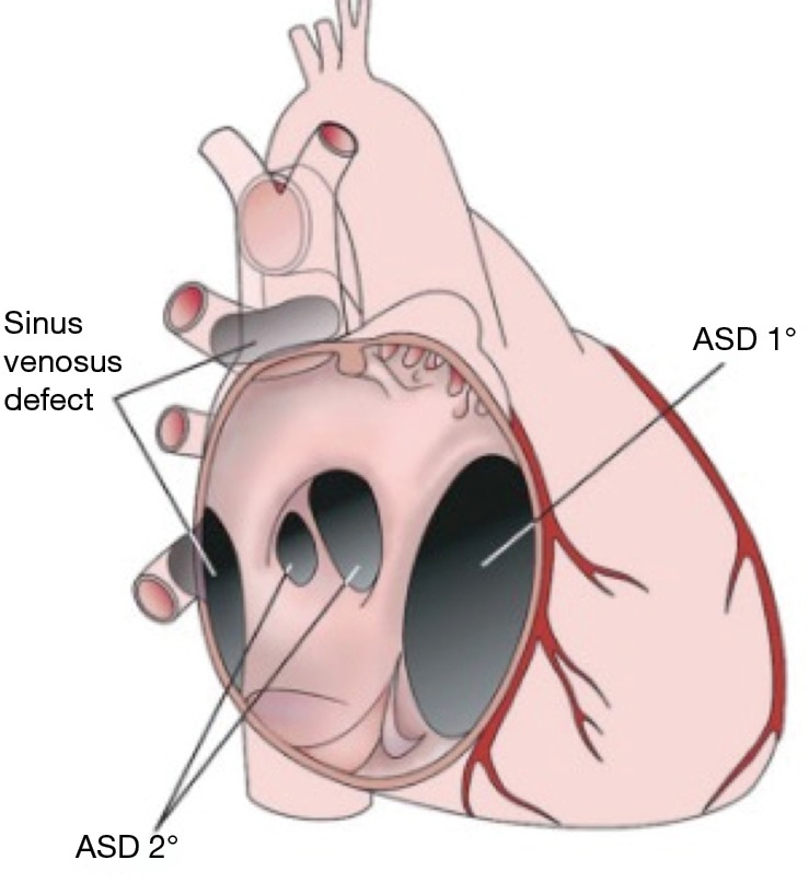
Types of ASD. ASD, atrial septal defect.
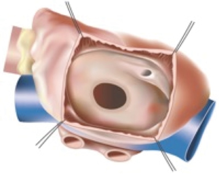
Secundum ASD. ASD, atrial septal defect.
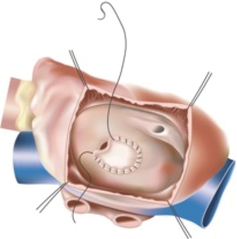
Patch closure of secundum ASD. ASD, atrial septal defect.
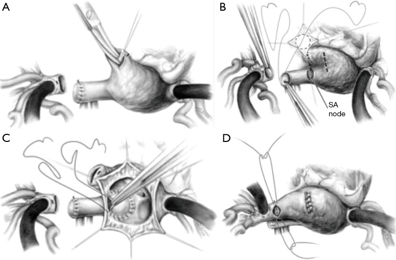
Warden technique for sinus venosus defect. (A) Division of SVC above anomalous pulmonary venous entry. (B) Patch closure of SVC above pulmonary vein entrance, opening in right atrial appendage and excision of pectinate muscle bundles. (C) Patch/baffle closure of ASD, connecting SVC with attached anomalous pulmonary venous drainage to the left atrium. (D) Right atrial appendage to SVC anastomosis and direct closure of atriotomy. ASD, atrial septal defect; SVC, superior vena cava.
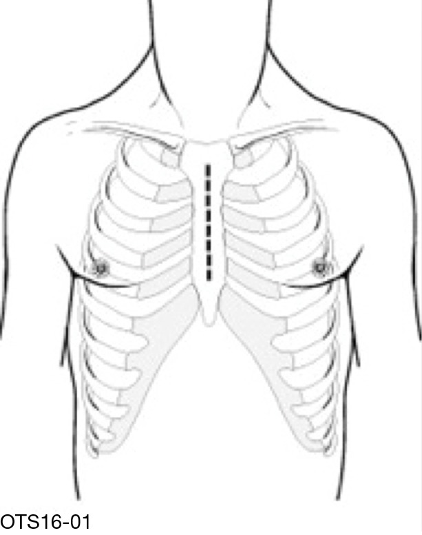
Median sternotomy.
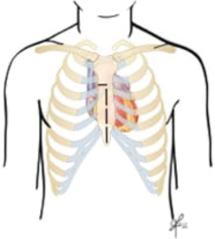
Partial sternotomy.
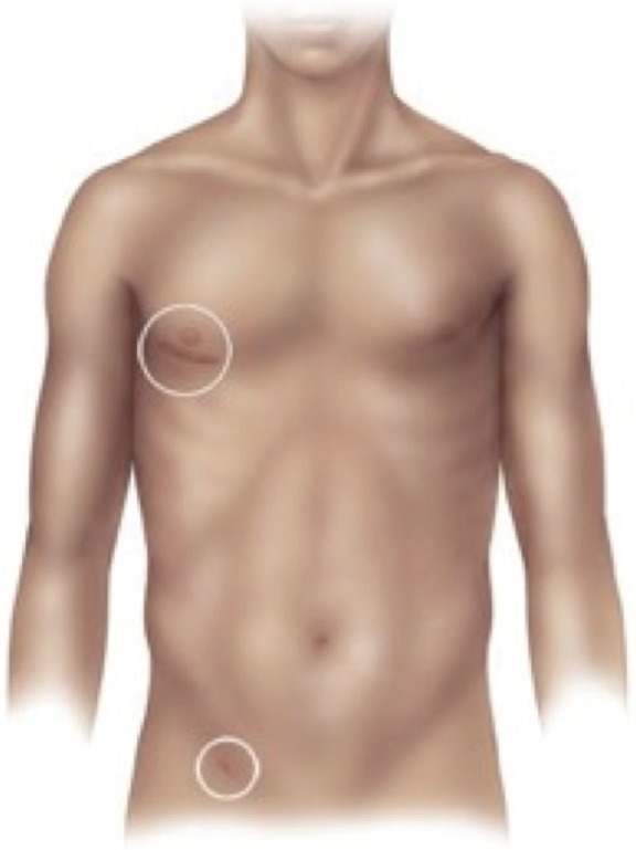
Right anterior thoracotomy and femoral incision.
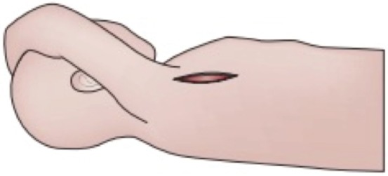
Vertical axillary incision.
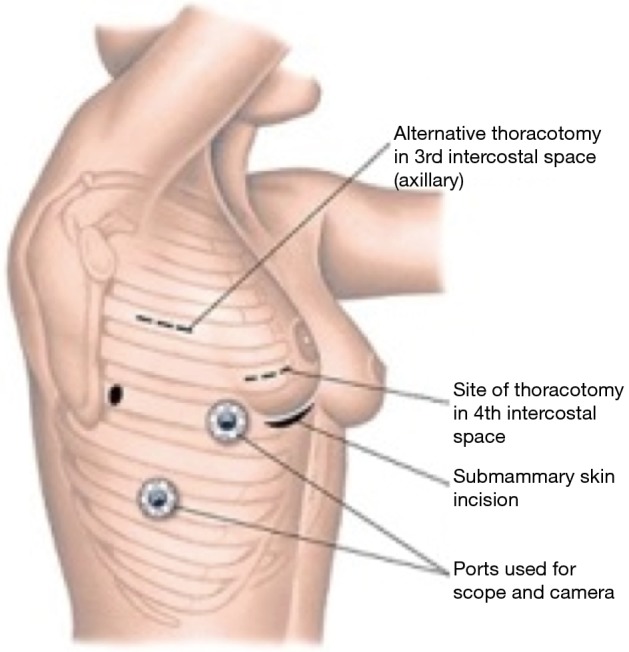
VATS incisions. VATS, video-assisted thoracoscopic surgery.
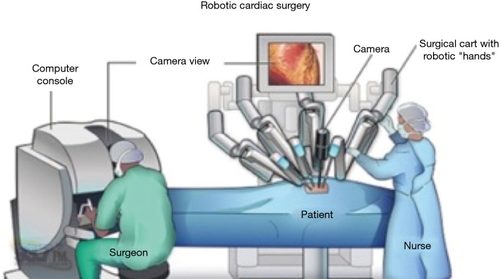
Robotic approach.
References
-
- Lewis FJ, Taufic M. Closure of atrial septal defects with the aid of hypothermia; experimental accomplishments and the report of one successful case. Surgery 1953;33:52-9. - PubMed
-
- Nagendran J, Habib HF, Kiaii B, et al. Minimally invasive endoscopic repair of atrial septal defects via right minithoracotomy. Multimed Man Cardiothorac Surg 2016;2016. - PubMed
Publication types
LinkOut - more resources
Full Text Sources