Unexpected Complication Ten Years after Initial Treatment: Long-Term Report and Fate of a Maxillary Premolar Rehabilitation
- PMID: 30305964
- PMCID: PMC6164210
- DOI: 10.1155/2018/3287965
Unexpected Complication Ten Years after Initial Treatment: Long-Term Report and Fate of a Maxillary Premolar Rehabilitation
Abstract
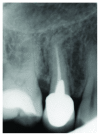
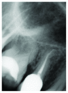
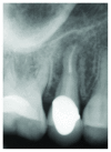
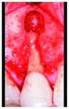
Full-coverage restorations represent a well-known rehabilitation strategy for compromised posterior teeth; in the last years, new ceramic materials like zirconia have been introduced and widely adopted for the prosthetic management of molar and premolar areas. A long-term follow-up of a maxillary premolar rehabilitation using a veneered zirconia crown is presented; after ten years of uneventful clinical service of the tooth-restoration complex, a serious complication-namely, a vertical root fracture (VRF)-occurred. An extended time lapse (9 years) between the end of restorative procedures and development of symptoms due to VRF has been observed. On the other hand, a complete functional and esthetic integrity of the zirconia crown (without chippings or crack development) is documented along the follow-up period. Due to periodontal breakdown and severity of fracture, the premolar was extracted. The illustrations of our late failure, aetiological factors, and available data on the literature regarding VRF are addressed. Patients and clinicians should be aware of potential occurrences of some long-term, serious complications when dealing with previously treated and/or structurally weakened teeth. The development of a VRF might be unexpected and might occur many years after the end of tooth rehabilitation, despite adoption of contemporary restorative protocols and techniques.
Figures


Similar articles
-
Zirconia-based versus metal-based single crowns veneered with overpressing ceramic for restoration of posterior endodontically treated teeth: 5-year results of a randomized controlled clinical study.J Dent. 2017 Oct;65:56-63. doi: 10.1016/j.jdent.2017.07.004. Epub 2017 Jul 21. J Dent. 2017. PMID: 28736293 Clinical Trial.
-
Zirconium dioxide based dental restorations. Studies on clinical performance and fracture behaviour.Swed Dent J Suppl. 2011;(213):9-84. Swed Dent J Suppl. 2011. PMID: 21919311
-
Clinical study to evaluate the wear of natural enamel antagonist to zirconia and metal ceramic crowns.J Prosthet Dent. 2015 Sep;114(3):358-63. doi: 10.1016/j.prosdent.2015.03.001. Epub 2015 May 16. J Prosthet Dent. 2015. PMID: 25985742
-
Prevalence of vertical root fracture as the reason for tooth extraction in dental clinics.Clin Oral Investig. 2015 Jul;19(6):1405-9. doi: 10.1007/s00784-014-1357-4. Epub 2014 Nov 15. Clin Oral Investig. 2015. PMID: 25398363
-
The Effect of Resin Bonding on Long-Term Success of High-Strength Ceramics.J Dent Res. 2018 Feb;97(2):132-139. doi: 10.1177/0022034517729134. Epub 2017 Sep 6. J Dent Res. 2018. PMID: 28876966 Free PMC article. Review.
References
Publication types
LinkOut - more resources
Full Text Sources

