

The Tentorium Cerebelli: A Comprehensive Review Including Its Anatomy, Embryology, and Surgical Techniques
- PMID: 30305987
- PMCID: PMC6168052
- DOI: 10.7759/cureus.3079
The Tentorium Cerebelli: A Comprehensive Review Including Its Anatomy, Embryology, and Surgical Techniques
Abstract
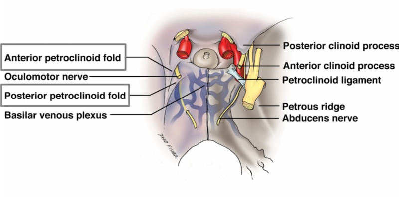
The tentorium cerebelli functions as a partition, dispelling the burden of weight from supratentorial structures upon inferior brain matter. Clinicians and neurosurgeons, when assessing pathological findings, should have knowledge regarding the tentorium cerebelli anatomy. This work of literature is a comprehensive review of the tentorium cerebelli, including its anatomy, embryology, and clinical and surgical implications. The evolutionary pattern demonstrates sequential stages to higher mammalian lineage. An understanding of the complexity of the neurovascular structures and the anatomy of the tentorium cerebelli is crucial for surgical procedures by neurosurgeons.
Keywords: dural sinus; embryology; incisura; tentorial notch; tentorium cerebelli.
Conflict of interest statement
The authors have declared that no competing interests exist.
Figures


References
-
- The cranial dura mater: a review of its history, embryology, and anatomy. Adeeb N, Mortazavi MM, Tubbs RS, Cohen-Gadol AA. Child Nerv Syst. 2012;28:827–837. - PubMed
-
- Adult cranial dura I: intrinsic vessels. Shukla V, Hayman LA, Ly C, Fuller G, Taber KH. J Compu Assist Tomo. 2002;26:1069–1074. - PubMed
-
- The tentorium in axial section. I. normal CT appearance and non-neoplastic pathology. Naidich TP, Leeds NE, Kricheff II, Pudlowski RM, Naidich JB, Zimmerman RD. Radiology. 1977;123:631–638. - PubMed
-
- The comparative anatomy and phylogeny of the tentorium cerebelli. Klintworth GK. Anat Rec. 1968;160:635–641. - PubMed
-
- Herniation of the brain. Meyer A. Arch Neuro Psychiatr. 1920;4:387–400.
Publication types
LinkOut - more resources
Full Text Sources