Review
. 2018 Sep 6:15:35-49.
doi: 10.1016/j.jot.2018.08.004. eCollection 2018 Oct.

Osteoporotic vertebral endplate and cortex fractures: A pictorial review

Affiliations
Review

Osteoporotic vertebral endplate and cortex fractures: A pictorial review

Yì Xiáng J Wáng et al. J Orthop Translat. .

Abstract

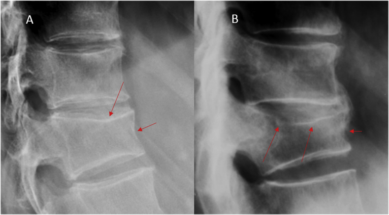
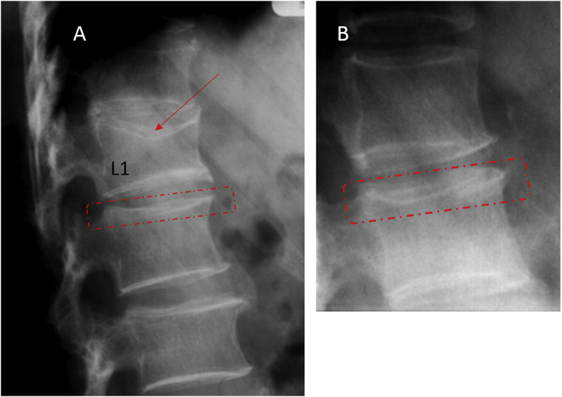
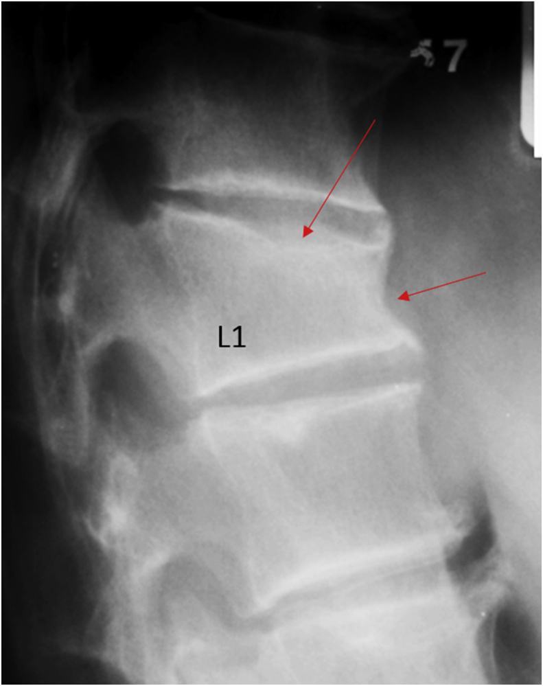
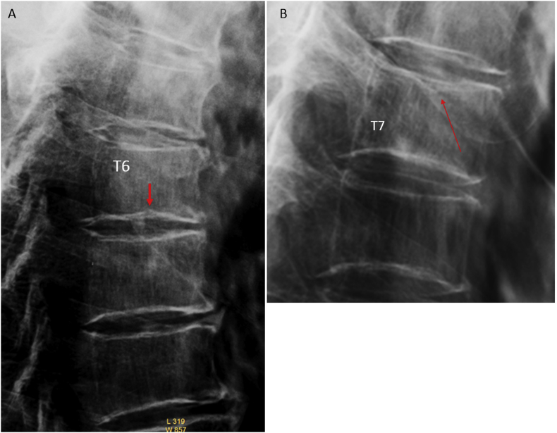
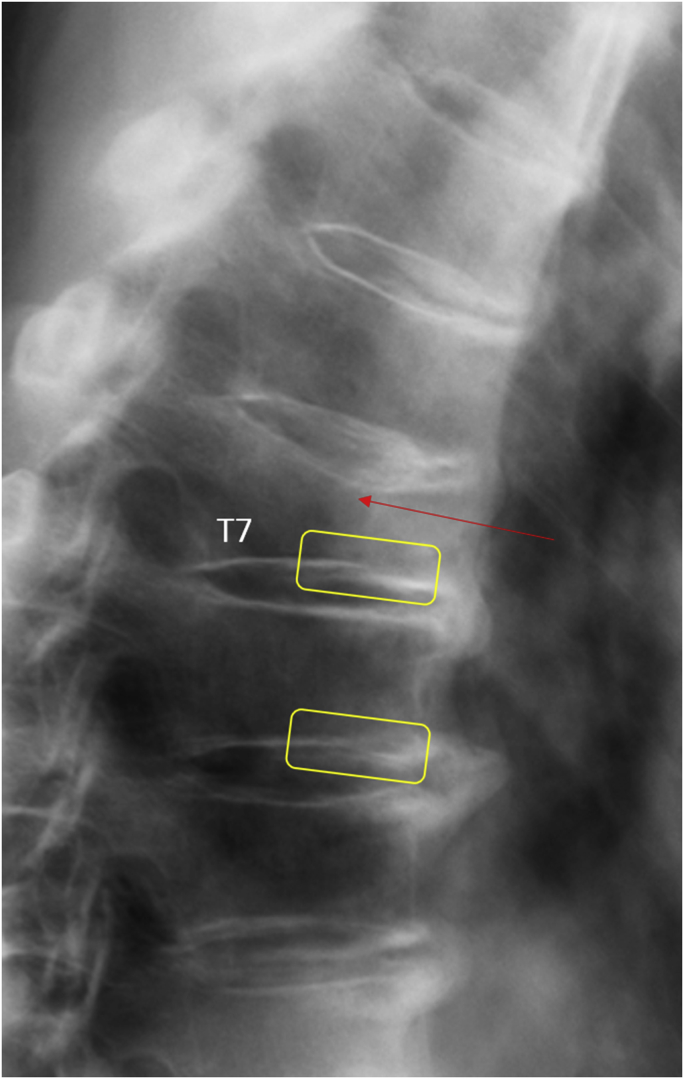
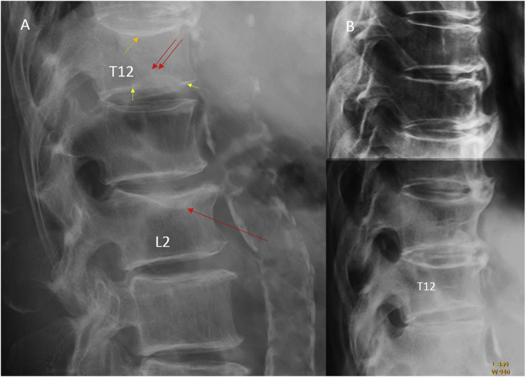
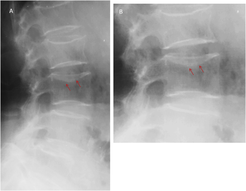
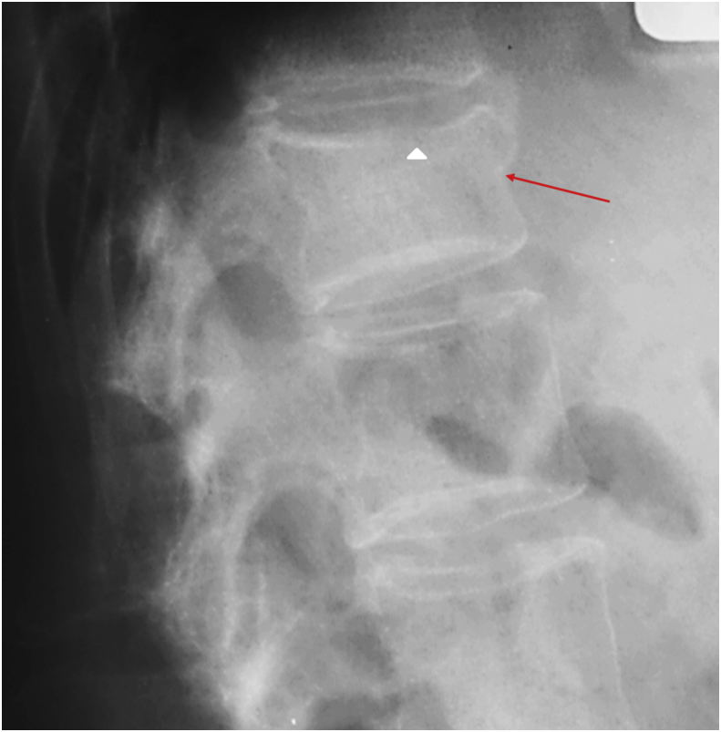
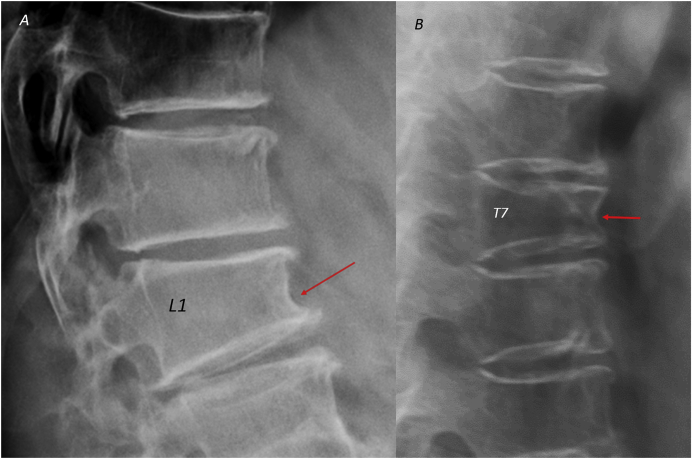
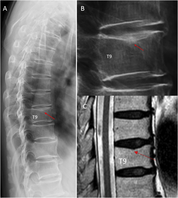
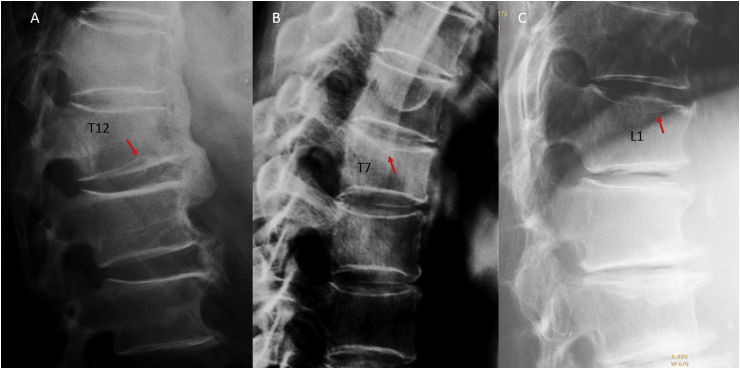
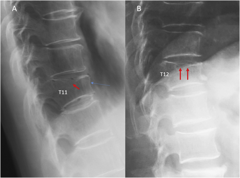
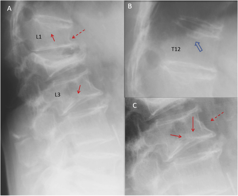
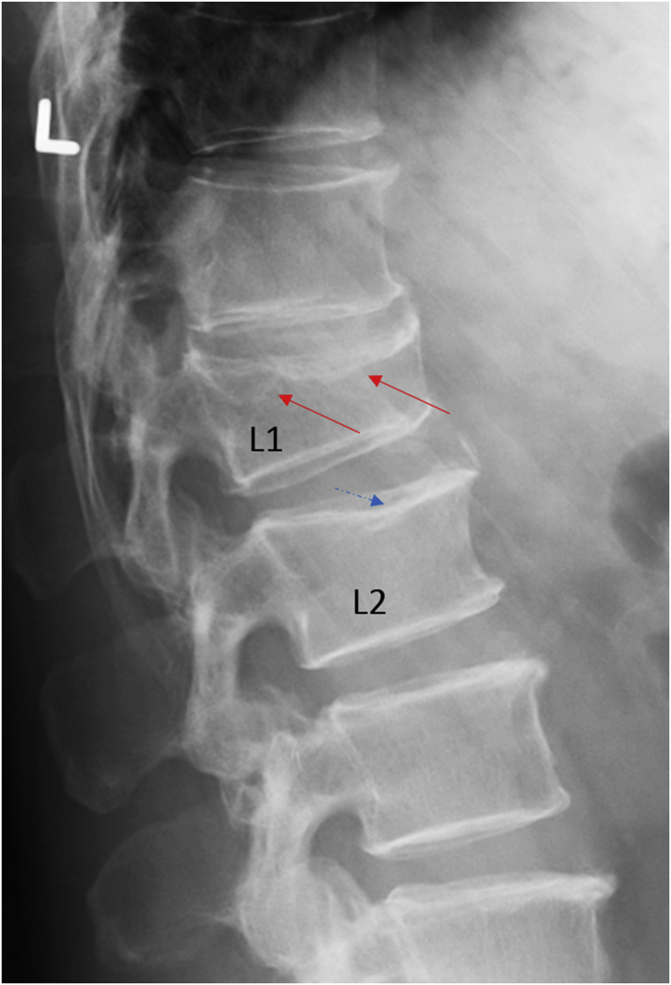
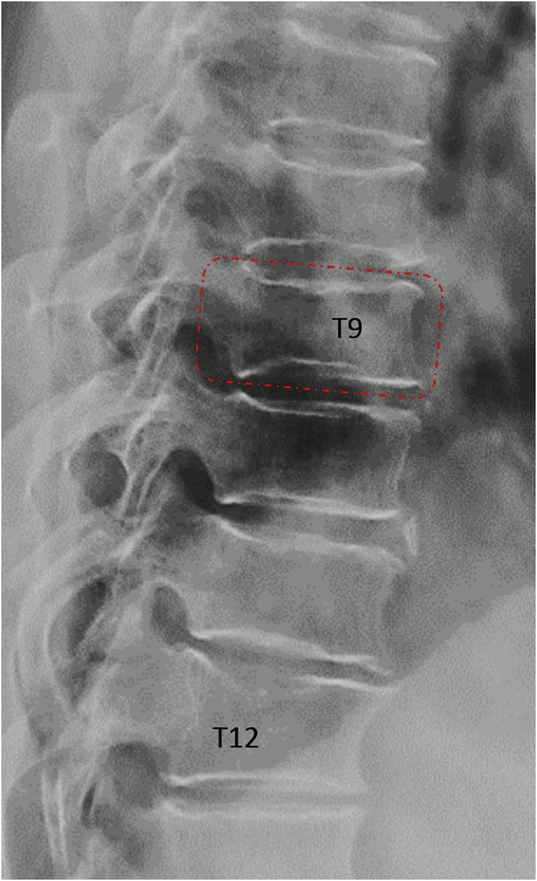
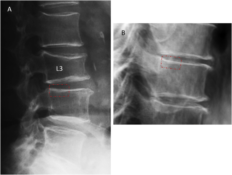
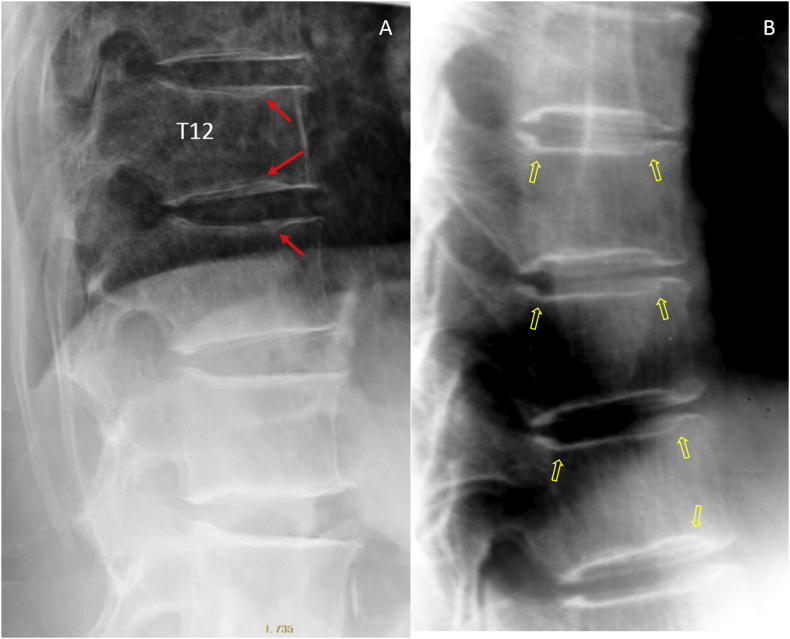
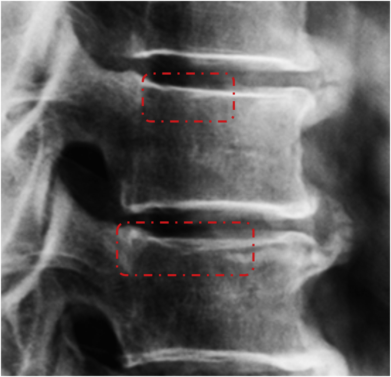
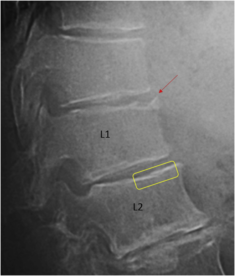
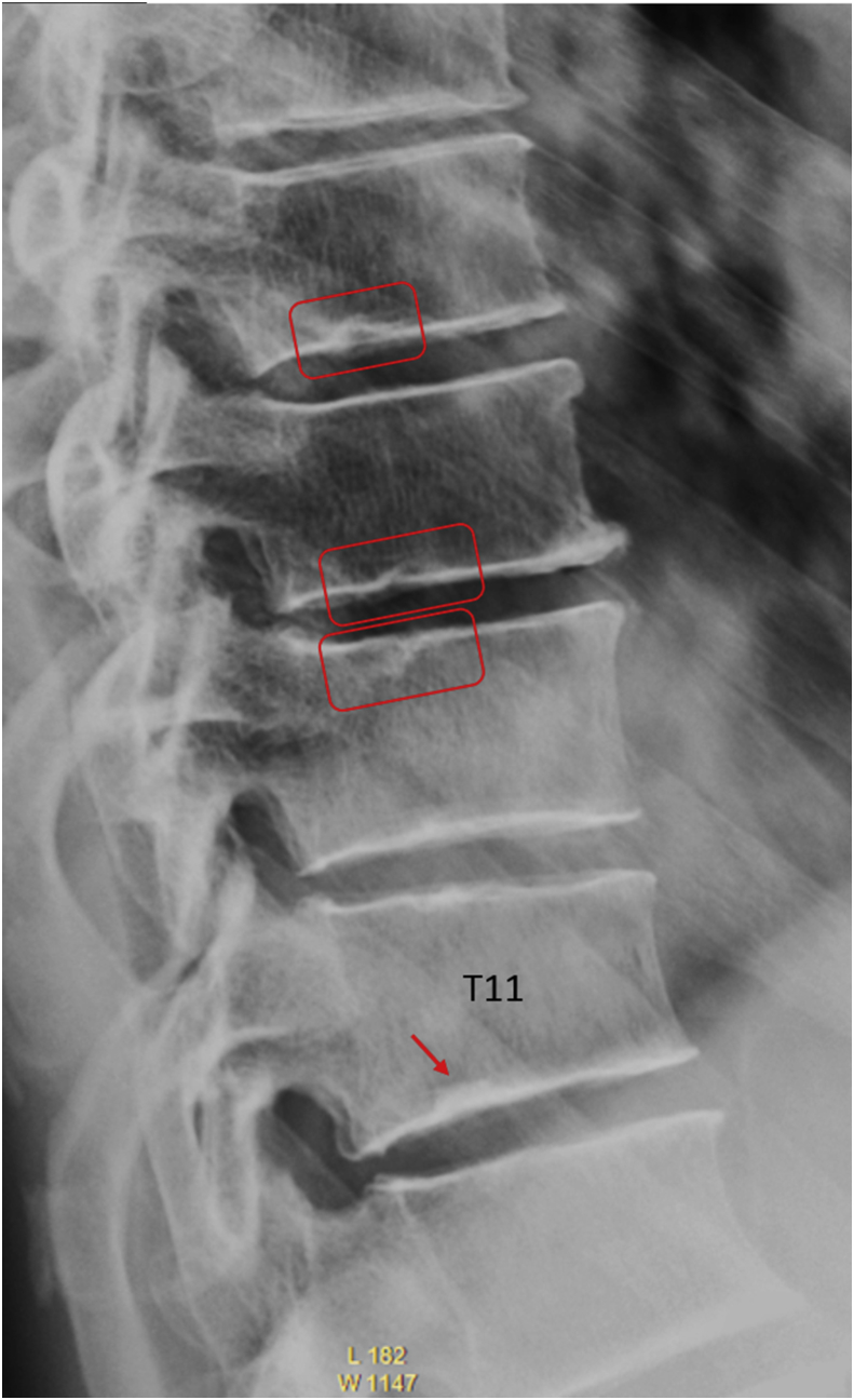
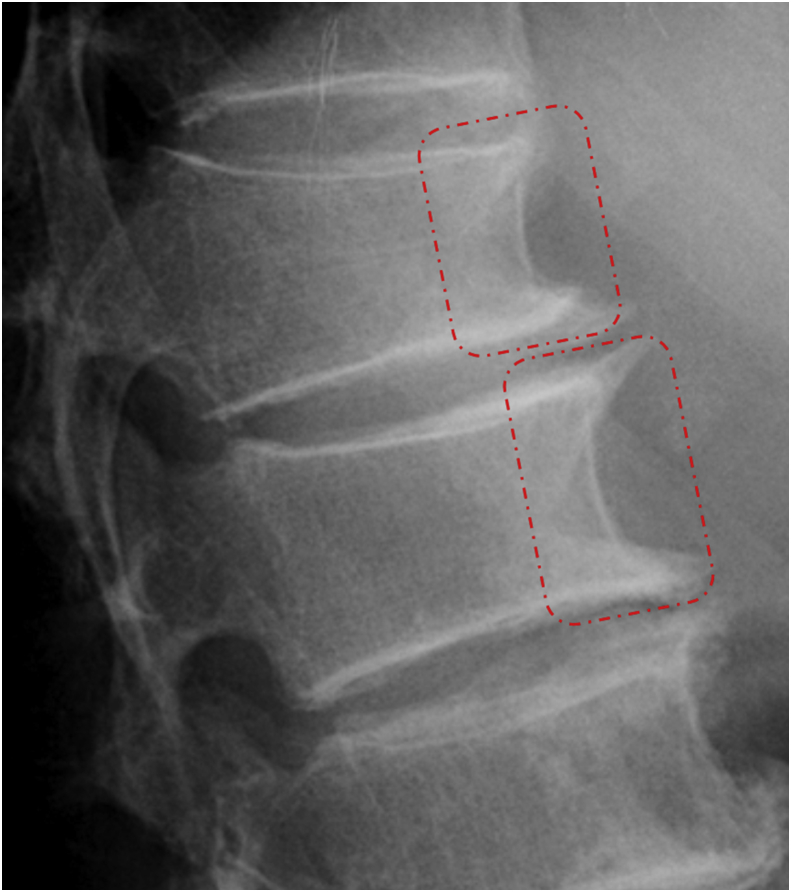
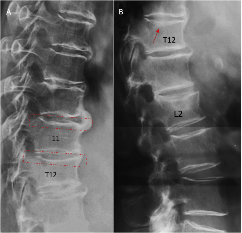
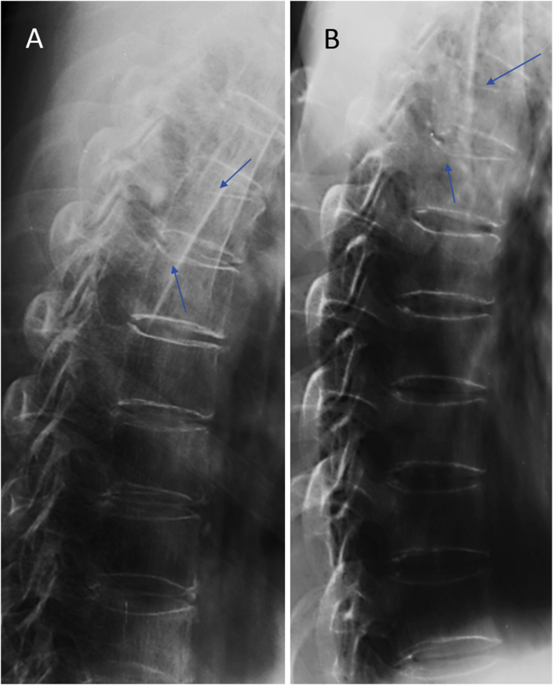
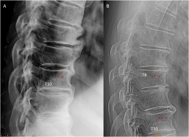
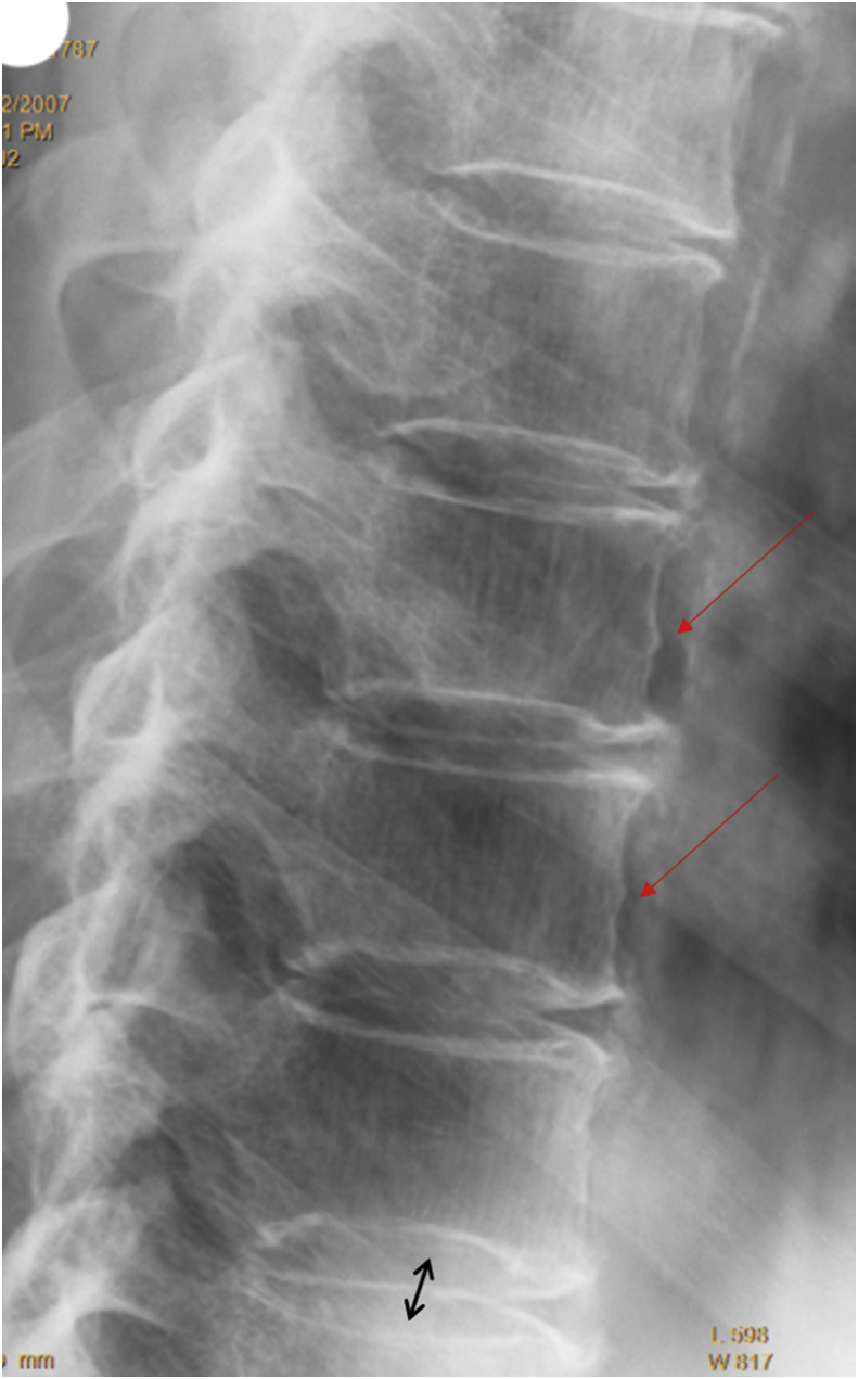
Despite years' research, the radiographic criteria for osteoporotic vertebral fracture and its grading remain debated. The importance of identifying vertebral endplate/cortex fracture (ECF) is being recognised; however, evaluation of osteoporotic ECF requires training and experience. This article aims to serve as a teaching material for radiologists/physicians or researchers to evaluate osteoporotic ECF. Emphasis is particularly dedicated to identifying ECF that may not be associated with apparent vertebral body collapse. We suggest a combined approach based on standardised radiologic evaluation by experts and morphometry measurement is the most appropriate approach to detect and classify osteoporotic vertebral fractures.

The translational potential: A good understanding of radiologic anatomy of vertebrae and fracture signs of endplate/cortex are essential for spine fragility fracture assessment.

Keywords: Endplate; Normal variants; Osteoporosis; Osteoporotic fractures; Radiograph.


Figures

Figure 1
Figure 1
(A) T12 upper endplate fracture (long arrow) and anterior cortex mild buckling (short arrow), with SQ-VD Grade 2; (B) T12 vertebra Grade 3 SQ-VD (wedge fracture) with buckling of the anterior cortex (short arrow) and upper endplate fracture (long arrow). Note that there is no fracture for T12 lower endplate. SQ-VD = semiquantitative vertebral deformity.
Figure 2
Figure 2
(A) L1 upper concave endplate fracture (long arrow) and Grade 2 SQ-VD. L2 upper endplate is normal (dotted square) which may be misdiagnosed as endplate fracture. This is due to the projection of endplate ring; note the parallel multilayered endplate lines and that there is no depression; (B) Radiograph from another subject demonstrates a similar appearance of upper endplate (without fracture). SQ-VD = semiquantitative vertebral deformity.
Figure 3
Figure 3
L1 vertebra Grade 3 SQ-VD and upper endplate fracture (long arrow) and anterior cortex mild buckling (short arrow). SQ-VD = semiquantitative vertebral deformity.
Figure 4
Figure 4
(A) T6 lower endplate fracture with broken endplate line and slight depression at its centre. There is no SQ-VD; (B) T7 upper endplate fracture and slight depression at its centre. There is no SQ-VD. SQ-VD = semiquantitative vertebral deformity.
Figure 5
Figure 5
T7 vertebra Grade 2 SQ-VD with upper endplate fracture (long arrow). The T7 lower endplate is without endplate fracture (the same appearance is also seen at T8 lower endplate). SQ-VD = semiquantitative vertebral deformity.
Figure 6
Figure 6
(A) L2 upper endplate fracture (red long arrow); A depression of T12 lower endplate is seen (double red arrow). The T12 lower endplate depression is symmetrically looking; the depressed endplate is of low density and there are angles (short yellow arrows) between the depressed endplate and nondepressed endplate. Our reading is that it could be a Schmorl's node (preferred diagnosis) or an endplate fracture. T12 upper endplate shows arc-shaped endplates (orange short arrow, commonly called “bean-can” appearance). This can usually be confirmed with comparing the endplates of upper adjacent vertebrae as shown in (B).
Figure 7
Figure 7
(A) L3 upper endplate fracture with depression (red arrows), and borderline Grade 0 SQ-VD (19% height reduction); (B) magnified image of the involved vertebra. SQ-VD = semiquantitative vertebral deformity.
Figure 8
Figure 8
L1 vertebra with Grade 2 SQ-VD. The arrow indicates anterior cortex fracture, and the triangle indicates ambiguous upper endplate fracture. SQ-VD = semiquantitative vertebral deformity.
Figure 9
Figure 9
(A) L1 vertebra anterior cortex buckling (arrow) and Grade 2 SQ-VD (anterior height loss approximately 38%). There is no endplate fracture observed for this vertebra; (B) T7 vertebra Grade 2 SQ-VD with upper endplate fracture and anterior cortex buckling (arrow). Note that the layered appearance of T8 upper endplate does not suggest fracture (see Figure 2). SQ-VD = semiquantitative vertebral deformity.
Figure 10
Figure 10
(A) T9 upper endplate fracture with slight depression at its centre; (B) magnified image of the involved vertebra. (C) A follow-up T2-weighted MR image 10 years later shows upper endplate central depression, with vertebral body homogenous fatty bone marrow signal. MR = magnetic resonance; SQ-VD = semiquantitative vertebral deformity.
Figure 11
Figure 11
(A) T12 vertebra Grade 2 SQ-VD and lower endplate fracture; (B) T7 vertebra Grade 1 SQ-VD and upper endplate fracture; (C) L1 vertebra Grade 2 SQ-VD and upper endplate fracture. SQ-VD = semiquantitative vertebral deformity.
Figure 12
Figure 12
(A) T11 vertebra Grade 2 SQ-VD with upper endplate depression (red arrow) and mild buckling of the anterior cortex (blue arrow, note that the vertebral borders have been marked with pen); (B) T12 vertebra Grade 2 SQ-VD and upper endplate fracture with depression and broken endplate line (double red arrows). SQ-VD = semiquantitative vertebral deformity.
Figure 13
Figure 13
Radiographs in (A), (B) and (C) are from the same patient. (A) L1 shows upper endplate fracture (red arrow) and anterior cortex buckling (red dotted arrow). L2 vertebra is collapsed (Grade 3 SQ-VD); (B) T12 Schmorl's node (blue arrow); (C) zoomed-in image of L3. There is lower endplate fracture (red arrows); however, L3 anterior cortex should not be read as buckling (red dotted arrow). SQ-VD = semiquantitative vertebral deformity.
Figure 14
Figure 14
L1 vertebra Grade 3 SQ-VD and upper endplate wavy fracture. L2 upper endplate ambiguous fracture (differential diagnosis of Schmorl's node cannot be excluded, or endplate fracture and Schmorl's node may coexist). SQ-VD = semiquantitative vertebral deformity.
Figure 15
Figure 15
T12 vertebra wedge deformity (Grade 2 SQ-VD) and upper endplate fracture. Note that both upper and lower endplates of T9 vertebra are without fracture (dotted square). SQ-VD = semiquantitative vertebral deformity.
Figure 16
Figure 16
(A) L3 upper and lower endplates fracture and SQ-VD grade 2. Radiographs (A) and (B) are from two subjects. The upper endplates in (A) and (B) marked by the red dotted rectangles are without fracture (a potential pitfall). SQ-VD = semiquantitative vertebral deformity.
Figure 17
Figure 17
(A) Red arrows indicate parts of endplate without fracture (potential pitfalls); (B) more typical type of these appearances (yellow arrows).
Figure 18
Figure 18
Dotted squares indicate normal appearance of upper endplate which may be misread as a fracture (potential pitfall).
Figure 19
Figure 19
Anterior upper part of L1 vertebra shows anterior marginal cartilage node (congenital anomaly, red arrow). L2 upper endplate is without fracture (yellow square); also see Figure 17B.
Figure 20
Figure 20
Vertebral T11 physiological (developmental) wedging and Schmorl's node (red arrow) at the lower endplate. Red rectangles indicate multiple Schmorl's nodes.
Figure 21
Figure 21
Dotted squares indicate intact anterior cortex (a potential pitfall). T12 (associated with upper dotted square) has Grade 2 SQ-VD with anterior height loss of 33%. Osteophytes are noted. SQ-VD = semiquantitative vertebral deformity.
Figure 22
Figure 22
T12 upper endplate shows fracture (lower square in A); this is better shown in B (arrow) and confirmed by comparing the upper endplates of T11 (upper square in A), T12 and L1. L2 Grade 2 SQ-VD upper endplate fracture and anterior cortex buckling. SQ-VD = semiquantitative vertebral deformity.
Figure 23
Figure 23
Arrows indicate normal appearance of endplate in upper thoracic spine. Potential pitfall (due to X-ray projection) reading of endplate depression.
Figure 24
Figure 24
Arrows indicate anterior vascular grooves. Radiographs (A) and (B) are from two subjects.
Figure 25
Figure 25
Red long arrows indicate slight irregularity of vertebral anterior cortex, which should not be interpreted as cortex buckling. Black double-headed arrow indicates “bean-can” appearance due to X-ray projection.

Similar articles

Cited by

References

    1. Ross P.D. Clinical consequences of vertebral fractures. Am J Med. 1997;103:30S–42S. - PubMed
    1. Kado D.M., Browner W.S., Palermo L., Nevitt M.C., Genant H.K., Cummings S.R. Vertebral fractures and mortality in older women: a prospective study. Study of Osteoporotic Fractures Research Group. Arch Intern Med. 1999;159:1215–1220. - PubMed
    1. Morin S., Lix L.M., Azimaee M., Metge C., Majumdar S.R., Leslie W.D. Institutionalization following incident non-traumatic fractures in community-dwelling men and women. Osteoporos Int. 2012;23:2381–2386. - PubMed
    1. Cooper C., Atkinson E.J., Jacobsen S.J., O'Fallon W.M., Melton L.J., 3rd Population-based study of survival after osteoporotic fractures. Am J Epidemiol. 1993;137:1001–1005. - PubMed
    1. Nevitt M.C., Ettinger B., Black D.M. The association of radiographically detected vertebral fractures with back pain and function: a prospective study. Ann Intern Med. 1998;128(10):793–800. - PubMed