. 2018 Oct 3:9:145-153.
doi: 10.1016/j.bonr.2018.09.004. eCollection 2018 Dec.

Glucocorticoids cause mandibular bone fragility and suppress osteocyte perilacunar-canalicular remodeling

Affiliations

Glucocorticoids cause mandibular bone fragility and suppress osteocyte perilacunar-canalicular remodeling

A Sean Alemi et al. Bone Rep. .

Erratum in

Abstract

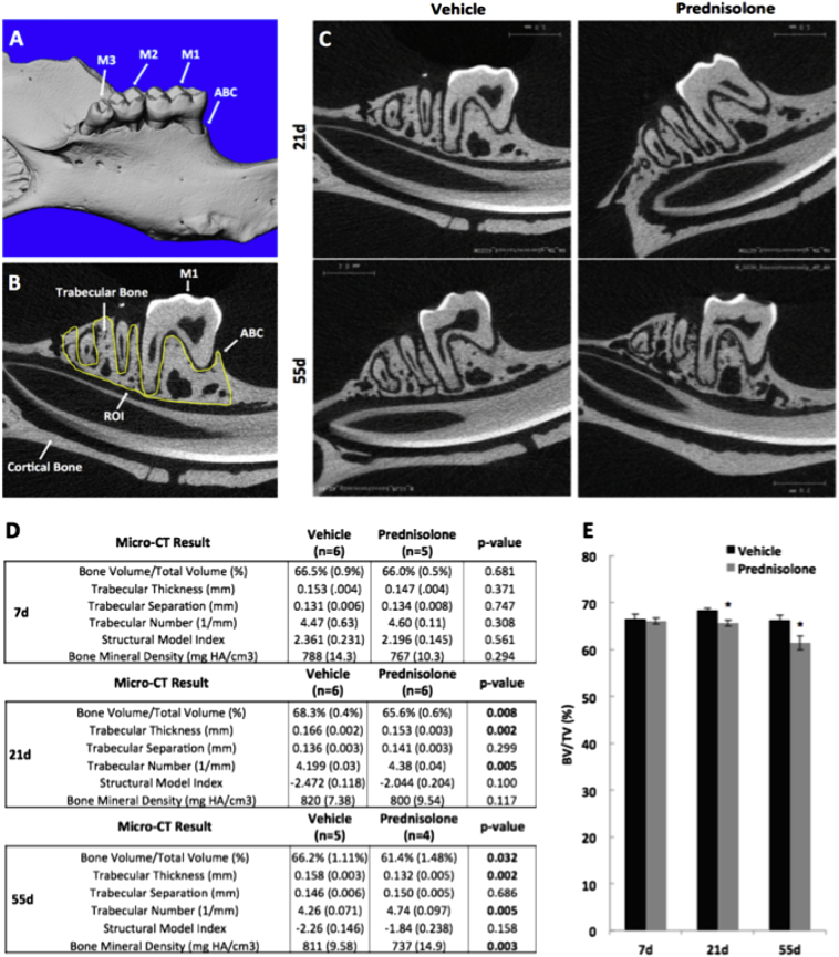
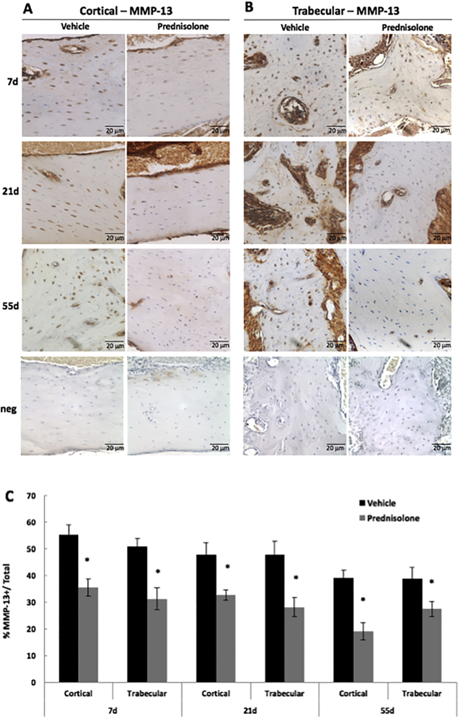
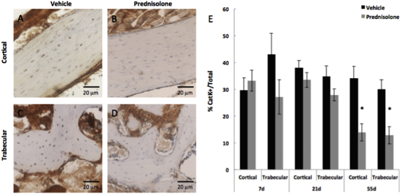
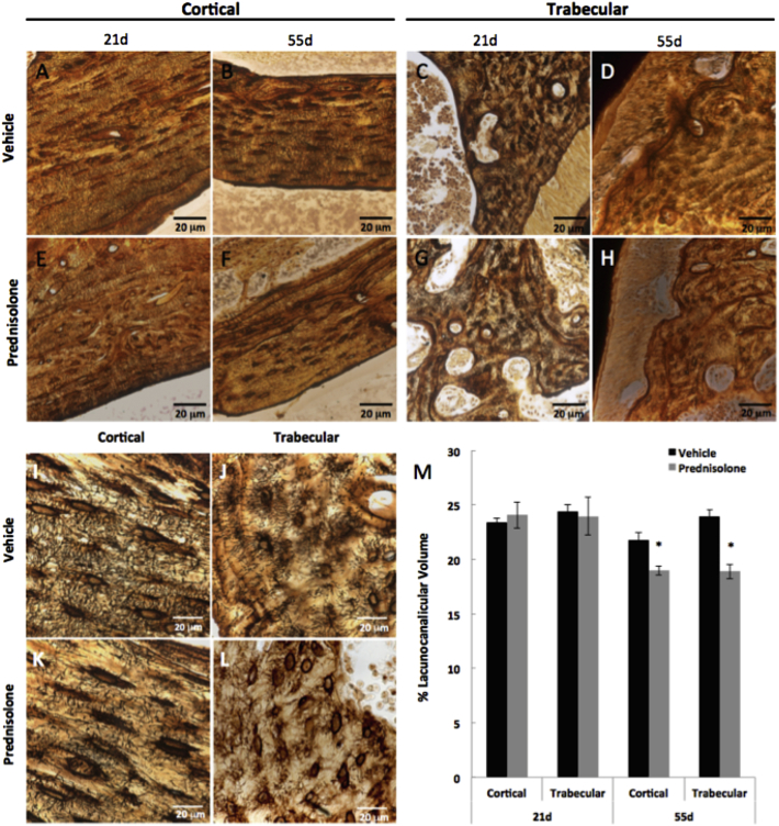
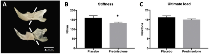
Osteocytes support dynamic, cell-intrinsic resorption and deposition of bone matrix through a process called perilacunar/canalicular remodeling (PLR). In long bones, PLR depends on MMP13 and is tightly regulated by PTH, sclerostin, TGFβ, and glucocorticoids. However, PLR is regulated differently in the cochlea, suggesting a mechanism that is anatomically distinct. Unlike long bones, the mandible derives from neural crest and exhibits unique susceptibility to medication and radiation induced osteonecrosis. Therefore, we sought to determine if PLR in the mandible is suppressed by glucocorticoids, as it is in long bone. Hemimandibles were collected from mice subcutaneously implanted with prednisolone or vehicle containing pellets for 7, 21, or 55 days (n = 8/group) for radiographic and histological analyses. Within 21 days, micro-computed tomography revealed a glucocorticoid-dependent reduction in bone volume/total volume and trabecular thickness and a significant decrease in bone mineral density after 55 days. Within 7 days, glucocorticoids strongly and persistently repressed osteocytic expression of the key PLR enzyme MMP13 in both trabecular and cortical bone of the mandible. Cathepsin K expression was significantly reduced only after 55 days of glucocorticoid treatment, at which point histological analysis revealed a glucocorticoid-dependent reduction in the lacunocanalicular surface area. In addition to reducing bone mass and suppressing PLR, glucocorticoids also reduced the stiffness of mandibular bone in flexural tests. Thus, osteocyte PLR in the neural crest-derived mandible is susceptible to glucocorticoids, just as it is in the mesodermally-derived femur, highlighting the need to further study PLR as a target of drugs, and radiation in mandibular osteonecrosis.

Keywords: Glucocorticoids; Mandible; Osteocyte; Perilacunar/canalicular remodeling.


Figures

Fig. 1
Fig. 1
Glucocorticoid treatment decreases mandibular bone volume. Micro-computed tomography (μCT) of hemi-mandibles from 8-week old male FVB mice treated with placebo or prednisolone for 21 or 55 days revealed significant differences in mandibular trabecular and cortical bone. (A) 3-Dimensional images were reconstructed showing the lingual surface of the mandible and the 1st, 2nd and 3rd molars (M1–3, respectively) and the alveolar bone crest (ABC). (B) Using 2-dimensional μCT images, we identified the region of interest (ROI, yellow line), which extends from the mesial aspect of the 1st molar (M1) to the distal aspect of M3. The height of the ROI is defined from the most distal aspect of any root to the alveolar bone crest (ABC). (D) Bone parameters measured by μCT at day 7, 21, and 55 include trabecular bone volume fraction/ total volume (BV/TV), trabecular thickness (Tb. Th), trabecular separation (Tb. Sp), trabecular number (Tb. N), structural model index (SMI), and bone mineral density. (E) Most notably BV/TV was significantly decreased between vehicle- and prednisolone-treated mice at day 21 and day 55. Error bars represent mean ± SEM; *P-value ≤ 0.05, n ≥ 4 compared to Placebo-treatment from Student's t-test. (For interpretation of the references to color in this figure legend, the reader is referred to the web version of this article.)
Fig. 2
Fig. 2
Glucocorticoids suppress MMP-13 expression by mandibular osteocytes. Immunohistochemistry (IHC) showed qualitatively diminished MMP-13 protein expression in osteocytes of prednisolone-treated mandibular cortical (A) and trabecular bone (B), relative to placebo-treated controls, after 7 days, 21 days, and 55 days. Neg denotes negative controls in which primary antibody was excluded from the IHC protocol. (C) The percent of MMP-13-postively stained osteocytes were normalized to the total number of osteocytes, revealing quantitatively fewer MMP-13-positive osteocytes in both trabecular and cortical bone after 7, 21, and 55 days of prednisolone-treatment. Scale bar = 20 μm (n ≥ 5), error bars indicate mean ± SEM, *P-value ≤ 0.05 compared to vehicle from Student's t-test.
Fig. 3
Fig. 3
Cathepsin K expression by mandibular osteocytes is suppressed by glucocorticoids after 55 days. Immunohistochemistry (IHC) for cathepsin K (Cat K) in vehicle- and prednisolone-treated cortical (A, B) and trabecular (C, D) bone after 55 days. Diminished positively stained osteocytes for Cat K were observed in both cortical and trabecular bone across all time points. (E) However, significant differences in the percentage of Cat K-positive osteocytes were only detectable at 55 days of treatment. Osteocytes positive for Cat K were quantified and normalized to total number of osteocytes in cortical or trabecular bone. Negative controls are shown in Fig. 2. Scale bar = 20 μm (n ≥ 5). Error bars indicate mean ± SEM, *P-value ≤ 0.05 compared to vehicle from Student's t-test.
Fig. 4
Fig. 4
Glucocorticoids disrupt the lacunocanalicular network of mandibular bone. Representative silver nitrate-stained histological sections of vehicle- and prednisolone-treated cortical bone after 21 days (A, E) and 55 days (B, F) and trabecular compartments after 21 days (C, G) and 55 days (D, H) respectively. High magnification images (100×) of 55 day vehicle- and prednisolone-treated bones portray the differences in lacunocanalicular network distribution and organization between cortical (I, K) and trabecular (J, L) compartments (scale bar = 20 μm). (M) Lacunocanalicular volume of osteocytes in cortical and trabecular bone is significantly decreased due to prednisolone treatment after 55 days. Scale bar = 20 μm (n ≥ 5). Error bars indicate mean ± SEM, *P-value ≤ 0.05 compared to vehicle from Student's t-test.
Fig. 5
Fig. 5
Glucocorticoid treatment causes severe fragility in mouse mandibles. HBSS-soaked hemi-mandibles were tested in three-point bending by positioning the buccal side in compression and the central loading point aligned at the midpoint of the molars. Hemi-mandibles from both groups consistently broke immediately posterior to the molars as shown by arrows on the buccal (top) and lingual (bottom) sides (A). In the glucocorticoid-treated mandibles, stiffness (B) was reduced by 17.2% relative to the placebo-treated controls after 35 days of treatment. Ultimate load (C) was not significantly affected by the prednisolone treatment after 35 days (N = 3 animals for placebo treatment, N = 4 animals for prednisolone treatment). Error bars indicate mean ± SEM; *P-value ≤ 0.05 compared to vehicle from Student's t-test.

Similar articles

Cited by

References

    1. Alliston T. Biological regulation of bone quality. Curr. Osteoporos. Rep. 2014;12(3):366–375. - PMC - PubMed
    1. Balooch G., Yao W., Ager J.W., Balooch M., Nalla R.K., Porter A.E., Ritchie R.O., Lane N.E. The aminobisphosphonate risedronate preserves localized mineral and material properties of bone in the presence of glucocorticoids. Arthritis Rheum. 2007;56(11):3726–3737. - PMC - PubMed
    1. Bélanger L.F., Jarry L., Uhthoff H.K. Osteocytic osteolysis in Paget's disease. Rev. Can. Biol. 1968;27(1):37–44. - PubMed
    1. Bellido T. Osteocyte-driven bone remodeling. Calcif. Tissue Int. 2014;94(1):25–34. - PMC - PubMed
    1. Bonewald L.F. The amazing osteocyte. J. Bone Miner. Res. 2011;26(2):229–238. - PMC - PubMed