. 2018 Oct 3;4(10):eaas9426.
doi: 10.1126/sciadv.aas9426. eCollection 2018 Oct.

Impact of parallel fiber to Purkinje cell long-term depression is unmasked in absence of inhibitory input

Affiliations

Impact of parallel fiber to Purkinje cell long-term depression is unmasked in absence of inhibitory input

Henk-Jan Boele et al. Sci Adv. .

Abstract

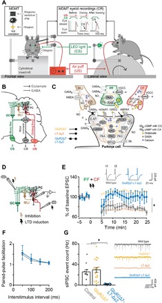
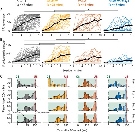
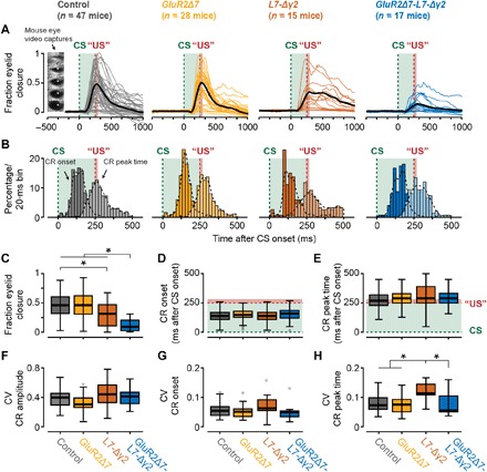
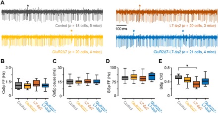
Pavlovian eyeblink conditioning has been used extensively to study the neural mechanisms underlying associative and motor learning. During this simple learning task, memory formation takes place at Purkinje cells in defined areas of the cerebellar cortex, which acquire a strong temporary suppression of their activity during conditioning. Yet, it is unknown which neuronal plasticity mechanisms mediate this suppression. Two potential mechanisms include long-term depression of parallel fiber to Purkinje cell synapses and feed-forward inhibition by molecular layer interneurons. We show, using a triple transgenic approach, that only concurrent disruption of both these suppression mechanisms can severely impair conditioning, highlighting that both processes can compensate for each other's deficits.


Figures

Fig. 1
Fig. 1. Eyeblink conditioning setup and confirmation that GluR2Δ7-L7-Δγ2 mice lack both PF-PC LTD and MLI-PC FFI.
(A) Mice were placed in a light- and sound-isolating chamber on a freely moving foam treadmill with their head fixed at a horizontal bar. The US consisted of a weak air puff, and the CS was a green light-emitting diode (LED) light. Eyelid movements were recorded with the magnetic distance measurement technique (MDMT). In vivo PC recordings were performed on the same treadmill system. (B) Neural circuits essential and sufficient for eyeblink conditioning, explained in detail in the main text. AIN, anterior interposed nucleus; GC, granule cells; IO, inferior olive; MLI, molecular layer interneurons; MN, motoneurons (N. III, VI, and VII); PC, Purkinje cell; PN, pontine nuclei. (C) Overview of the main known molecular mechanisms underlying PC plasticity. PF-PC LTD cascades are indicated with ocher arrows, MLI-PC cascades are indicated with brown arrows, and PF-PC long-term potentiation (LTP) and intrinsic plasticity are indicated with dashed arrows. GluR2Δ7 mice lack PF-PC LTD, L7-Δγ2 mice lack MLI-PC inhibition, and GluR2Δ7-L7-Δγ2 mice have disrupted both PF-PC LTD and MLI-PC inhibition. (D) Schematic overview of MLI-PC inhibition and PF-PC LTD induction experiments. (E) In contrast to controls, GluR2Δ7-L7-Δγ2 mice show no PF-PC LTD using an induction protocol that consisted of a 100-Hz PF stimulation in eight pulses, followed by a 110-ms delay of single CF activation at 1 Hz for 5 min. (F) Applying interstimulus intervals varying from 50 to 200 ms evoked similar levels of paired-pulse facilitation, indicating that baseline PF-PC presynaptic mediated neurotransmitter release is unaltered in GluR2Δ7-L7-Δγ2 mice. (G) GluR2Δ7-L7-Δγ2 mice show significantly less sIPSCs than controls, indicating that they indeed lack MLI-PC inhibition. For PF-PC LTD and sIPSCs, data of GluR2Δ7 and L7-Δγ2 mice are used with permission from Schonewille et al. (17) and Wulff et al. (19), respectively. AC, adenylyl cyclase; AMPAR, AMPA receptor (GluR2/3); CaMKII, Ca2+/calmodulin-activated kinase II; cAMP, cyclic adenosine monophosphate; cGMP, cyclic guanylate monophosphate; CRF, corticotropin-releasing factor; GABAA, GABA type A receptor; GABAB, GABA type B receptor; G.AP, GABAAR-associated protein; GC, guanylylcyclase; GluRδ2, glutamate receptor δ2 (GRID2); IP3, inositol trisphosphate; mGluR1, metabotropic glutamate receptor 1; NMDAR, N-methyl-d-aspartate receptor; NO, nitric oxide; PKA, cAMP-dependent protein kinase; PKC, protein kinase C; PKG, cGMP-dependent protein kinase; PLC, phospholipase C; PP1, protein phosphatase 1; PP2, protein phosphatase 2 (A+B); RAB; RAS-related protein RAB3A; SK, small conductance Ca2+-activated K+ channel; VGCC, voltage-gated Ca2+ channel. For complete statistics, we refer to table S1; *P < 0.05.
Fig. 2
Fig. 2. Acquisition of eyeblink CRs is severely impaired in GluR2Δ7-L7-Δγ2 mice, whereas GluR2Δ7 and L7-Δγ2 mice show normal or only mildly impaired conditioning.
(A and B) Mouse individual (colored) and average (black) learning curves per group showing the development of (A) CR percentage (B) and FECs (or “amplitude of eyelid closure”) over the 10 consecutive training sessions. GluR2Δ7-L7-Δγ2 mice have a significantly slower acquisition and lower CR percentage and FEC than the other three groups. L7-Δγ2 mice have a mild phenotype with a normal CR percentage but significantly lower FEC at the end of training. No significant difference could be established between controls and GluR2Δ7 mutants. (C) Peristimulus histogram plots with a Gaussian kernel density estimate (black dashed line) showing the distribution of CR onset (dark filled bars) and CR peak time (light filled bars) relative to CS and US onset in paired CS-US trials for sessions 1, 4, 7, and 10. Green dashed lines indicate CS onset, and red dashed lines denote US onset; light green and light red fill indicate CS and US duration, respectively. In all groups, there is a clear development in CR onset and peak time: There are no clearly preferred times in the CS-US interval at the start of training (session 1), but toward the end of training (sessions 7 and 10), CR onset values are centered around 100 to 125 ms after CS onset, and CR peak times are located around the onset of the expected US. In all panels, we refer to “CR” if only trials are included with an FEC > 0.1 and to “FEC” if all trials are included. For complete statistics, we refer to table S2; *P < 0.05.
Fig. 3
Fig. 3. Kinetic profile of eyeblink CRs during CS-only trials at the end of training.
(A) Mouse individual average (colored) and total average (black) eyeblink traces of CS-only trials in the probe session following acquisition. Mouse eye video captures show eyelid closure ranging from 0 (fully open) to 1 (fully closed). GluR2Δ7-L7-Δγ2 mice have significantly smaller FEC values than the other three groups. (B) Peristimulus histogram plots with a Gaussian kernel density estimate (black dashed line) showing the distribution of CR onset (dark filled bars) and CR peak time (light filled bars) relative to CS and US onset in paired CS-US trials for session 11 (probe). Green dashed lines indicate CS onset, and red dashed lines denote US onset; light green and light red fill indicate CS and US duration, respectively. (C) Mice carrying the GluR2Δ7-L7-Δγ2 mutation have significantly lower FEC values than the three other groups. In addition, L7-Δγ2 mice have significantly lower FEC values than control mice. FEC value is based on all CS-only trials in the probe session and represents the highest peak in the CS-US interval. (D and E) No significant difference was established for the average latency to CR onset and latency to CR peak. Both CR onset and CR peak time are based only on trials in which a CR was present (that is, FEC > 0.1 in the CS-US interval). Green dashed lines indicate CS onset, and red dashed lines denote US onset; light green and light red fill indicate CS and US duration, respectively. (F and G) No significant difference was established for CV values on CR amplitude and latency to CR onset. (H) As can be noted in the raw traces in (A), L7-Δγ2 mice have a more jittery CR peak time, which results in a significantly higher CV value compared to the other three groups. In all panels, we refer to “CR” if only trials with an FEC > 0.1 are included and to “FEC” if all trials are included. For complete statistics, we refer to table S2; *P < 0.05.
Fig. 4
Fig. 4. GluR2Δ7-L7-Δγ2 mice have no gross abnormalities in spontaneous firing behavior in vivo.
(A) Raw examples of in vivo PC recordings from lobules I to V for all four groups. For (B) complex spike (CoSp) firing frequencies, (C) simple spike pauses following complex spikes, and (D) simple spike (SiSp) firing frequencies, no differences were found between controls and any of the mutant groups. (E) Unlike L7-Δγ2 mice, GluR2Δ7-L7-Δγ2 mice have no significantly higher simple spike regularity, as shown by the lower CV2 value. For complete statistics, we refer to table S3; *P < 0.05.

References

    1. H. J. Boele, M. M. Ten Brinke, C. I. De Zeeuw, Classical conditioning of timed motor responses: Neural coding in cerebellar cortex and cerebellar nuclei, in The Neural Codes of the Cerebellum, D. H. Heck, Ed. (Academic Press, 2015), pp. 53–96.
    1. Freeman J. H., Steinmetz A. B., Neural circuitry and plasticity mechanisms underlying delay eyeblink conditioning. Learn. Mem. 18, 666–677 (2011). - PMC - PubMed
    1. Hesslow G., Correspondence between climbing fibre input and motor output in eyeblink-related areas in cat cerebellar cortex. J. Physiol. 476, 229–244 (1994). - PMC - PubMed
    1. Mostofi A., Holtzman T., Grout A. S., Yeo C. H., Edgley S. A., Electrophysiological localization of eyeblink-related microzones in rabbit cerebellar cortex. J. Neurosci. 30, 8920–8934 (2010). - PMC - PubMed
    1. Heiney S. A., Kim J., Augustine G. J., Medina J. F., Precise control of movement kinematics by optogenetic inhibition of Purkinje cell activity. J. Neurosci. 34, 2321–2330 (2014). - PMC - PubMed

Publication types

Substances

LinkOut - more resources