

Contrast-enhanced ultrasound patterns of hepatocellular adenoma: an Italian multicenter experience
- PMID: 30306412
- PMCID: PMC6531526
- DOI: 10.1007/s40477-018-0322-5
Contrast-enhanced ultrasound patterns of hepatocellular adenoma: an Italian multicenter experience
Abstract
Purpose: Hepatocellular adenoma (HCA) is a rare benign monoclonal neoplasm, recently categorized on genetic and histopathological basis into four subtypes with different biological behaviors. Since contrast-enhanced ultrasonography (CEUS) is nowadays a well-established technique for liver nodule characterization, the aim of our study was to assess CEUS features of HCAs to identify criteria that correlate with different HCA subtypes as compared to histopathologic examination and other imaging modalities.
Methods: We retrospectively analyzed data of patients with histology-proven HCA who underwent CEUS, computed tomography or magnetic resonance imaging (MRI) in seven different Italian ultrasound units.
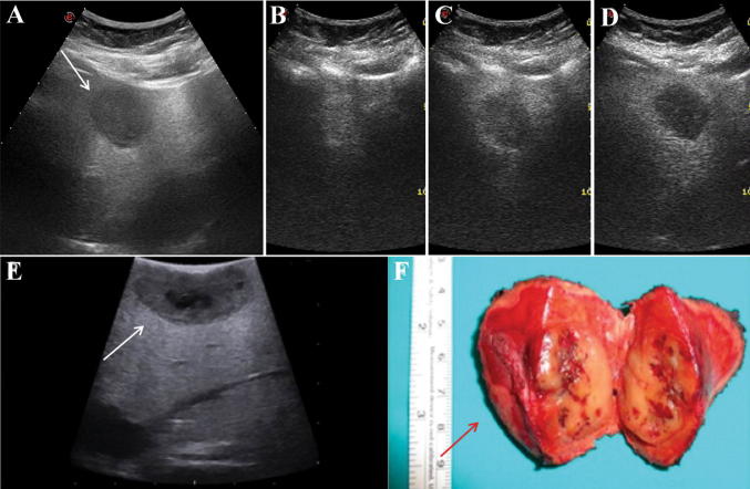
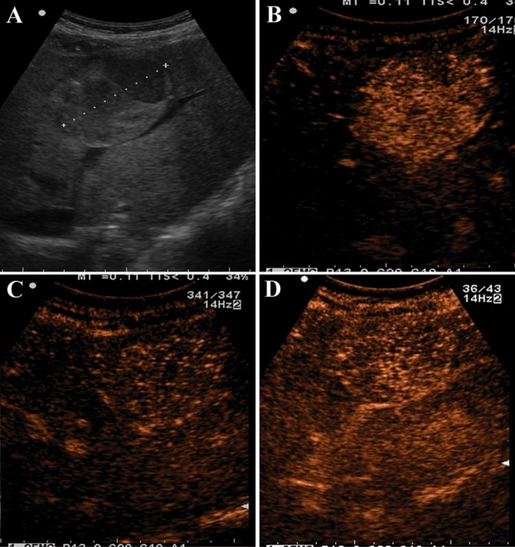
Results: The study enrolled 19 patients (16 females; 69% with concomitant/prior use of oral contraceptives): the mean size of all HCAs was 4.2 cm (range 1.6-7.1 cm); 14/19 had inflammatory HCAs (I-HCA), 1/19 β-catenin-activated HCA, and the others unclassified HCAs. On CEUS, during the arterial phase, all but one HCA displayed a rapid enhancement, with 89% of these showing centripetal and 11% centrifugal filling pattern, whereas during the portal and late venous phase 58% of HCA showed washout and the remaining 42% displayed persistent enhancement. In particular, among I-HCAs 7/14 showed no washout, 3/14 and 4/14 showed washout in the portal or late phase, respectively.
Conclusions: This dataset represents one of the few published experiences on HCAs and CEUS in Italy and shows that HCAs are hypervascularized in the arterial phase usually with a centripetal flow pattern and have a heterogeneous behavior in portal and late phase. In particular, occurrence of delayed washout on CEUS but not on MRI is frequently observed in the subtype of I-HCA.
Introduzione: L′adenoma epatico (HCA) rappresenta una rara neoplasia primitiva del fegato, recentemente classificata in quattro diversi sottotipi sulla base delle caratteristiche istopatologiche e del comportamento biologico. In considerazione dell’ampio e diffuso utilizzo dell’ecografia con mezzo di contrasto ecografico (CEUS) nella valutazione non-invasiva delle lesioni focali epatiche l’obiettivo di questo studio è stato quello di documentare in una casistica multicentrica le caratteristiche CEUS di lesioni focali epatiche già caratterizzate come HCA e di valutare le eventuali correlazioni con i diversi sottotipi istologici e con altre metodiche di imaging (CT/MRI).
Metodi: Sono stati raccolti retrospettivamente le informazioni su pazienti con diagnosi istologica di HCA sottoposti a CEUS e CT ± MR in sette diversi centri italiani di ecografia.
Risultati: Sono stati inclusi nello studio 19 pazienti con diagnosi istologica di HCA (16 donne; 69% con storia attuale e/o pregressa di utilizzo di farmaci estroprogestinici): 14/19 adenomi sottotipo “infiammatori” (IHCA), 1/19 β-catenin-activated HCA e i restanti erano HCA non classificabili. L’esame CEUS ha mostrato nella quasi totalità dei casi (18/19) un rapido enhancement arterioso di tipo centripeto (89%) o centrifugo (11%). Durante la fase portale e tardiva si è dimostrato un wash-out contrastografico rispettivamente nel 58% degli HCA; invece nel 42% dei rimanenti casi non è stato osservato wash-out in nessuna delle fasi contrastografiche. In particolare è stato evidenziato che nel sottotipo I-HCA 7/14 non presentavano washout in nessuna delle fasi contrastografiche, mentre 3/14 e 4/14 mostravano rispettivamente un washout nelle fasi portali o tardive.
Conclusioni: La nostra casistica rappresenta una delle poche esperienze italiane presenti in letteratura riguardo all’utilizzo della CEUS negli adenomi epatici, confermando l’aspetto di ipervascolarizzazione nella fase arteriosa (soprattutto con un flusso centripeto) ed il comportamento eterogeneo nelle fasi portali e tardive. In particolare, nel caso di I-HCA un comportamento contrastografico caratterizzato da washout in fase tardiva è frequente con l’utilizzo della CEUS ma non con l’utilizzo della MRI.
Keywords: Benign liver lesion; Contrast-enhanced ultrasound; Hepatocellular adenoma; Magnetic resonance imaging; Phenotype classification.
Conflict of interest statement
All authors declare no conflict of interest.
Figures
References
Publication types
MeSH terms
Substances
LinkOut - more resources
Full Text Sources
Medical
