. 2018 Oct;143 Suppl 2(Suppl 2):109-117.
doi: 10.1002/ijgo.12618.

Role of imaging in the routine management of endometrial cancer

Affiliations

Role of imaging in the routine management of endometrial cancer

Ming Yin Lin et al. Int J Gynaecol Obstet. 2018 Oct.

Abstract

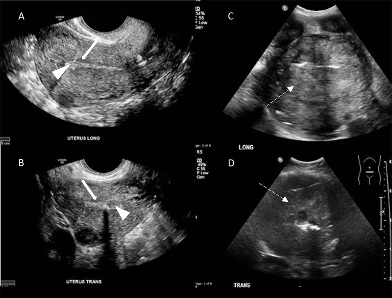
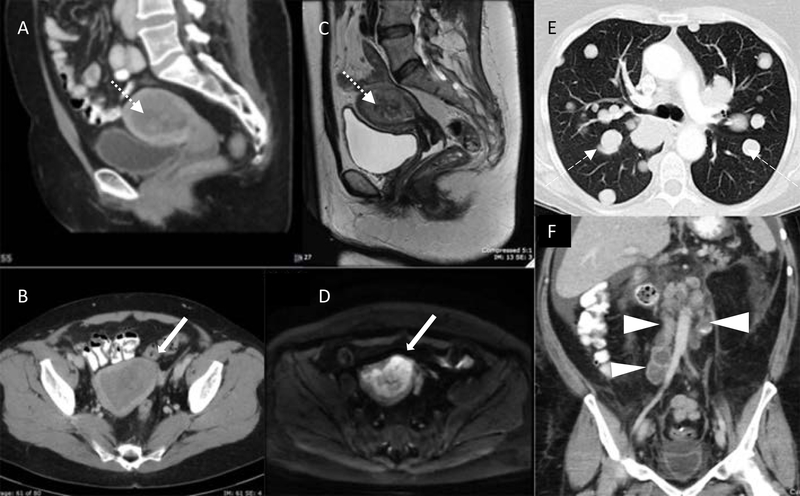
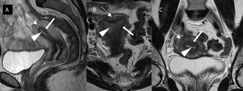
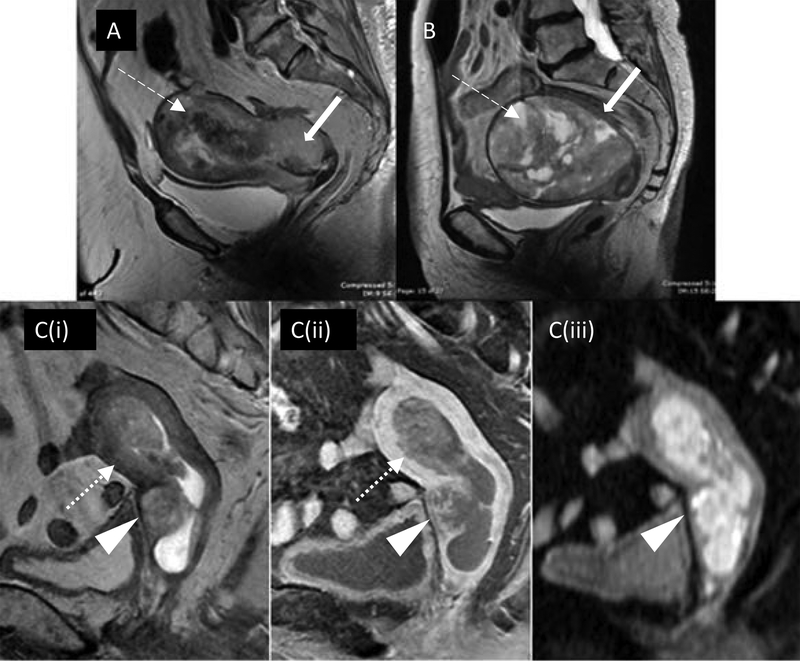
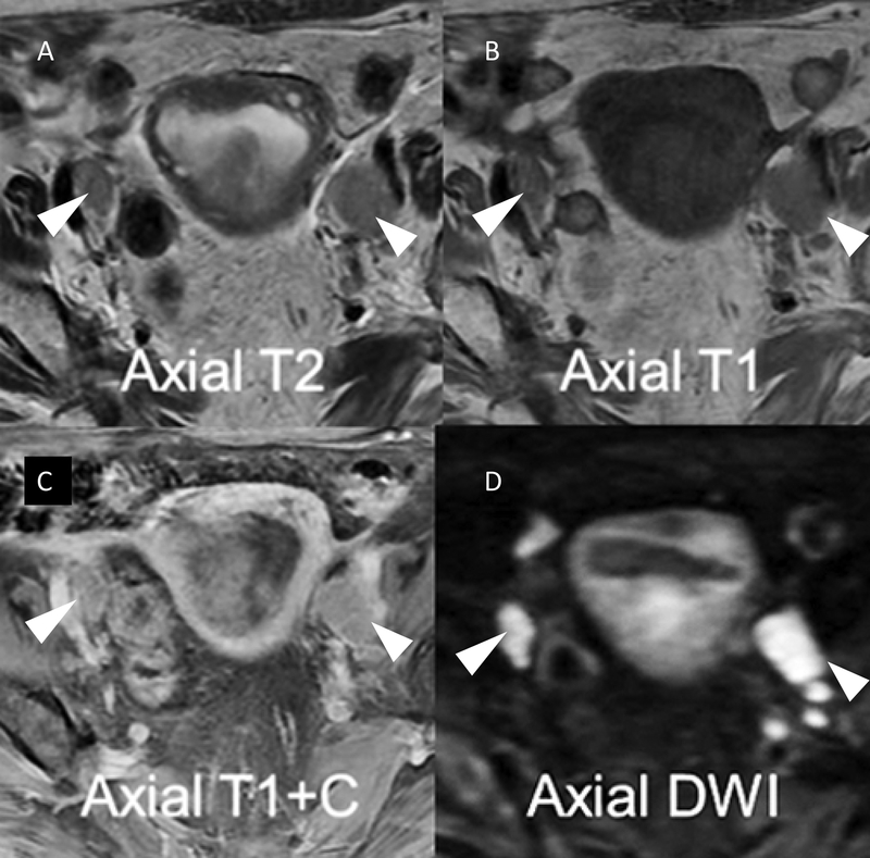
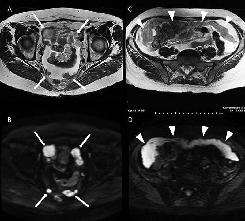
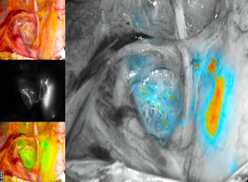
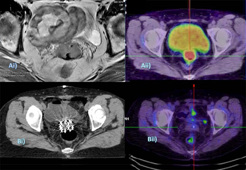
Endometrial cancer is the most common gynecologic cancer in women today. It is surgically staged, and while surgery is the primary treatment modality, the identification of disease extent-in particular extrauterine spread-prior to surgery is important to optimize treatment decision making. Ultrasound and MRI are useful for evaluating the extent of local disease, while CT and PET are used for detecting lymph node or distant metastases. Diffusion-weighted MRI has also been used for detecting small metastatic deposits in lymph nodes and omentum. Extrauterine soft tissue involvement can be detected by ultrasound, CT, MRI, and PET. Recently, intraoperative visualization techniques, such as sentinel lymph node mapping, are increasingly used to avoid extensive surgical staging without compromising treatment. Imaging is also used for planning adjuvant treatment and detection of postoperative residual disease in high-risk patients, monitoring and detecting recurrent disease, and in post-treatment surveillance of asymptomatic patients with high risk of relapse.

Keywords: Endometrial cancer; FIGO Cancer Report; MRI; PET; Radiology; Ultrasound.


Conflict of interest statement

Conflict of interest

The authors have no conflicts of interest to declare.

Figures

Figure 1
Figure 1
Ultrasound scans of a normal uterus. Transvaginal ultrasound scans of (A) sagittal plane (long) and (B) axial plane (trans) with normal, thin, uniform echogenic endometrium (arrowheads) and subendometrial hypoechoic halo (solid arrows). Transabdominal ultrasound scans of endometrial carcinoma in sagittal plane (C) and axial plane (D). Note heterogeneous echogenicity due to hemorrhage and necrosis (dashed arrows).
Figure 2
Figure 2
Computed tomography (A: sagittal plane, B: axial plane) and MRI (C: sagittal T2, D: axial diffusion-weighted) in the same patient with endometrial carcinoma distending the endometrial cavity (dotted arrows) and deep myometrial invasion (solid arrows). E: Axial CT chest with multiple “cannonball” pulmonary metastases (dashed arrows). F: Coronal CT abdomen demonstrating multiple para-aortic necrotic lymph node metastases (arrowheads)
Figure 3
Figure 3
Normal zonal anatomy on T2-weighted MRI in (A) sagittal, (B) axial, and (C) coronal planes demonstrating high signal (bright) endometrium (arrowheads), low signal (darker) junctional zone or inner myometrium (solid white arrows), and intermediate signal (grey) outer myometrium (dotted arrows).
Figure 4
Figure 4
Deep myometrial invasion on MRI: (A) Sagittal T2 demonstrating obvious deep myoinvasion with heterogeneous tumor obliterating the normal uterine zonal anatomy (dashed arrow) and invading cervical stroma (solid arrow). (B) Pitfall of MRI myometrial invasion assessment: large polypoid tumor stretches and thins the myometrium (dashed arrow) with age-related loss of normal zonal anatomy (solid arrow), pathology confirming no evidence of deep myoinvasion. (C) Multiparametric MRI improving staging accuracy: equivocal depth of myoinvasion and cervical stromal involvement on sagittal T2 (Ci). Dynamic T1 fat saturated post contrast (Cii) shows disruption of subendometrial stripe at anterior midbody of uterus (dotted arrow) confirming myoinvasion. Cervical mucosal enhancement preserved posteriorly but disrupted anteriorly (arrowhead) confirming stromal invasion. Diffusion-weighted imaging (Ciii) highlights tumor extent and deep cervical stromal invasion but absence of bladder wall invasion (arrowhead).
Figure 5
Figure 5
Lymph node evaluation on MRI is enhanced by the combination of high resolution T2 (A) and T1(B) anatomical imaging and functional sequences including post contrast T1 fat saturated (C) and diffusion-weighted imaging increasing nodal conspicuity and improving morphological assessment (arrowheads).
Figure 6
Figure 6
Peritoneal metastatic nodular deposits (solid arrows) demonstrated on axial T2 (A) and diffusion-weighted imaging (B) sequences in the same patient. Omental cake (arrowheads) on axial T2: (C) intermediate signal: grey and prominent restricted diffusion on diffusion-weighted imaging; (D): high signal: bright.
Figure 7
Figure 7
Example of sentinel lymph node mapping with indocyanine green cervical injection using the PINPOINT system. The right external iliac sentinel lymph node is seen medial to the external iliac vessels, and ventral to the internal iliac vessels. Also demonstrated are lymphatic trunks from the right paracervix crossing over the obliterated umbilical ligament on the way to the sentinel lymph node.
Figure 8
Figure 8
Use of imaging in diagnosis, treatment planning, and restaging of recurrent endometrial cancer: (Ai) MRI (axial) shows the vaginal vault mass encased within loops of bowel; and (Aii) Corresponding slice on pre-radiotherapy PET-CT, performed to confirm isolated recurrence. (Bi) Radiotherapy planning CT with interstitial implant needles in situ; and (Bii) Six-month post-treatment restaging PET-CT.

References

    1. Siegel RL, Miller KD, Jemal A. Cancer statistics, 2018. CA Cancer J Clin 2018;68(1):7–30. - PubMed
    1. Talhouk A, McAlpine JN. New classification of endometrial cancers: the development and potential applications of genomic-based classification in research and clinical care. Gynecol Oncol Res Pract 2016;3:14. - PMC - PubMed
    1. Dijkhuizen FP, Mol BW, Brolmann HA, Heintz AP. The accuracy of endometrial sampling in the diagnosis of patients with endometrial carcinoma and hyperplasia: a meta-analysis. Cancer 2000;89:1765–1772. - PubMed
    1. Abdaal A, Mushtaq Y, Khasati L, Moneim J, Khan F, Ahmed H, et al. Post-menopausal bleeding - Is transvaginal ultrasound a useful first-line investigation in tamoxifen users? Post Reprod Health 2018:2053369118755190. - PubMed
    1. Creasman WT, Morrow CP, Bundy BN, Homesley HD, Graham JE, Heller PB. Surgical pathologic spread patterns of endometrial cancer. A Gynecologic Oncology Group Study. Cancer 1987;60(8 Suppl):2035–2041. - PubMed