Review
. 2019 Feb;29(1):1-12.
doi: 10.1089/nat.2018.0747. Epub 2018 Oct 16.

Cell-Penetrating Peptide Conjugates of Steric Blocking Oligonucleotides as Therapeutics for Neuromuscular Diseases from a Historical Perspective to Current Prospects of Treatment

Affiliations
Review

Cell-Penetrating Peptide Conjugates of Steric Blocking Oligonucleotides as Therapeutics for Neuromuscular Diseases from a Historical Perspective to Current Prospects of Treatment

Michael J Gait et al. Nucleic Acid Ther. 2019 Feb.

Abstract

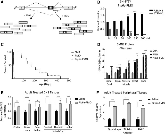
The review starts with a historical perspective of the achievements of the Gait group in synthesis of oligonucleotides (ONs) and their peptide conjugates toward the award of the 2017 Oligonucleotide Therapeutic Society Lifetime Achievement Award. This acts as a prelude to the rewarding collaborative studies in the Gait and Wood research groups aimed toward the enhanced delivery of charge neutral ON drugs and the development of a series of Arg-rich cell-penetrating peptides called Pip (peptide nucleic acid/phosphorodiamidate morpholino oligonucleotide [PNA/PMO] internalization peptides) as conjugates of such ONs. In this review we concentrate on these developments toward the treatment of the neuromuscular diseases Duchenne muscular dystrophy and spinal muscular atrophy toward a platform technology for the enhancement of cellular and in vivo delivery suitable for widespread use as neuromuscular and neurodegenerative ON drugs.

Keywords: conjugate; neuromuscular; oligonucleotide; peptide; therapeutic.


Conflict of interest statement

M.J.G., M.J.A.W., and C.G. are cofounder shareholders of PepGen, a company dedicated to the peptide-based enhancement of delivery of therapeutic oligonucleotide analogs. All other authors declare no conflicts of interest.

Figures

<b>FIG. 1.</b>
FIG. 1.
Nucleotide analogs used by the Gait group for studies of steric block ON activities in comparison to 2′-MOE nucleotides used in the Ionis research studies. ON, oligonucleotide.
<b>FIG. 2.</b>
FIG. 2.
Development of novel Pip sequences starting from the CPP Penetratin. These include on the left side R6-Penetratin (R6-Pen), Pip1, which was the initial peptide disulfide linked to PNA 705 for splicing redirection, Pip2a and Pip2b, the initially protease stabilized peptides, as well as the more recent Pip5e and Pip6a. PMO sequences and their linkage functionalities are shown on the right side. R6-Pen and early Pip were conjugated to an anti-TAR PNA or a splice-correcting PNA705 through a disulfide linkage, whereas later Pip were either stably thioether linked to PNA705 or amide linked to PMO for exon skipping in DMD mdx cells. CPP, cell-penetrating peptide; DMD, duchenne muscular dystrophy.
<b>FIG. 3.</b>
FIG. 3.
(A) The HIV-1 trans-activation assay; (B) inhibition of HIV-1 TAR RNA trans-activation by R6Pen-PNA as measured by the reduction of firefly luciferase activity.
<b>FIG. 4.</b>
FIG. 4.
(A) Splicing redirection assay by ON 705 to block aberrant splicing in HeLa 705 cells, and (B) fold increases in luminescence for R6-Pen-PNA705 and Pip1-PNA705 (note the different scales on the Y axis).
<b>FIG. 5.</b>
FIG. 5.
(A) The exon skipping assay using PNA ONs in mdx mouse cells in culture. The cross denotes a premature termination signal, introduced as a result of a point mutation in exon 23 of the dystrophin gene, which gives rise to a truncated nonfunctional protein; (B, C) intramuscular delivery in the mdx mouse showing (B) results as seen by RT-PCR for naked PNA, [R-Ahx-R]4-PNA, Pip1-PNA, Pip2a-PNA, and Pip2b-PNA showing the increased exon skipping levels for Pip2a and Pip2b-PNAs, and (C) number of dystrophin-positive fibers in immunohistochemistry staining for the same conjugates. Asterisks refer to the use of five mice rather than three mice for other experiments. Figures reproduced from reference [24].
<b>FIG. 6.</b>
FIG. 6.
(A) Exon skipping in mdx cells in culture Pip2b-PNA, Pip5e-PNA, and Pip5e-PMO and (B) percentage of dystrophin positive fibers seen for Pip5-PMO conjugates following systemic injections into mdx mice, reproduced from reference [34].
<b>FIG. 7.</b>
FIG. 7.
Sequences of Pip6-PMO conjugates, cardiac muscle splicing, and protein restoration following systemic administration of conjugates. A single 12.5 mg/kg systemic injection of peptide-PMO was administered to mdx mice, and tissues were harvested 2 weeks later. (A) A list of names and sequences, including rationale for synthesis of the peptides used in the study. (B) Percentage Δ23 exon skipping determined by q-RT-PCR, (C) dystrophin protein restoration determined by western blot, and (D) dystrophin immunohistochemistry staining quantified relative to control laminin counter-stain in heart tissue of C57BL10 and mdx untreated and mdx treated mice. Relative intensity values for each region of interest (120 regions) are plotted. (E) Representative dystrophin staining images for C57BL10, mdx untreated, and some Pip6 conjugates. Inverted (Pip6a) and scrambled (Pip6f) hydrophobic regions show homogenous dystrophin expression compared to the shortened (Pip6c) peptide. R = arginine; X = aminohexanoic acid; B = beta-alanine. qRT-PCR, quantitative real time-PCR.
<b>FIG. 8.</b>
FIG. 8.
(A) Table of exemplar CPP sequences of Pip 7, 8, and 9 series of CPP-PMOs. (B, C) Splice-switching activity in tibialis anterior of mdx mice following a single 12.5 mg/kg intravenous administration as measured by (B) quantitative RT-PCR of exon 23 skipping levels and (C) dystrophin protein restoration as assessed by western blot. R–arginine; X–aminohexanoic acid; B–beta-alanine.
<b>FIG. 9.</b>
FIG. 9.
(A) Schematic of human SMN2 alternative splicing of exon 7. The majority of transcripts produced from SMN2 exclude exon 7 (Δ7SMN2). Antisense oligonucleotides targeting intron 7 intron splice suppressor element (ISSN1) sterically hinder the binding of trans-splice repressor proteins to generate more exon 7-included transcripts (FLSMN2). (B) Dose-dependent expression of increasing FLSMN2 and decreasing Δ7SMN2 transcripts observed in cultured neuroblastoma SH-SY5Y cells following treatment with Pip6a-PMO. Q-PCR expression was normalized to “total SMN2” transcripts represented by amplification of exons 2a and 2b. Data are represented as the mean ± SEM. **P ≤ 0.005, ***P ≤ 0.0005, Student's t-test relative to untreated expression. (C) Direct comparison of systemic PMO versus Pip6a-PMO administration in severe SMA pups. Untreated SMA pups survive a median of 12 days before reaching their humane end point. At 10 μg/g, PMO on its own did not improve survival (median 11.5 days), while 10 μg/g Pip6a-PMO significantly enhanced survival (mean of 196.4 ± 114.9; median 167 days) (P ≤ 0.0001 Log-rank Mante-Cox). (D) Survival reflective of expression of SMN2 protein in spinal cord, brain, and skeletal muscle 7 days postadministration. Protein analyzed by western blots for human SMN and mouse β-tubulin. Data represented as mean ± SEM. *P ≤ 0.05, **P ≤ 0.01, ***P ≤ 0.001 Student's t-test in comparison to untreated SMA mice. (E, F) Pip6a-PMO administration into unaffected adult mice harboring the human SMN2 allele (Smn1tm1Hung/WT; SMN2tg/tg). Tissues from the (E) brain and spinal cord and (F) peripheral skeletal muscles and liver were harvested 7 days post 18 mg/kg Pip6a-PMO administration. In all tissues, FLSMN2 expression was significantly increased over saline treated mice. Data represented by mean ± SEM. *P ≤ 0.005, **P ≤ 0.001, ***P ≤ 0.0001, Student's t-test in comparison to saline treated mice [43]. SMA, spinal muscular atrophy.

References

    1. Lundin KE, Gissberg O. and Smith CI. (2015). Oligonucleotide therapies: the past and the present. Hum Gene Ther 26:475–485 - PMC - PubMed
    1. Shen X. and Corey DR. (2018). Chemistry, mechanism and clinical status of antisense oligonucleotides and duplex RNAs. Nucleic Acids Res 46:1584–1600 - PMC - PubMed
    1. Crooke ST. (2017). Molecular mechanisms of antisense oligonucleotides. Nucleic Acid Ther 27:70–77 - PMC - PubMed
    1. Järver P, O'Donovan L. and Gait MJ. (2014). A chemical view of oligonucleotides for exon skipping and related drug applications. Nucleic Acid Ther 24:37–47 - PMC - PubMed
    1. Aartsma-Rus A, Straub V, Hemmings R, Haas M, Schlosser-Weber G, Stoyanova-Beninska V, Mercuri E, Muntoni F, Sepodes B, Vroom E, Balabanov P. (2017). Development of exon skipping therapies for Duchenne Muscular Dystrophy: a critical review and a perspective on the outstanding issues. Nucleic Acid Ther 27:251–259 - PMC - PubMed

MeSH terms