Review
. 2019;20(2):150-156.
doi: 10.1080/15384047.2018.1523852. Epub 2018 Oct 11.

Diagnosis of NUT carcinoma of lung origin by next-generation sequencing: case report and review of the literature

Affiliations
Review

Diagnosis of NUT carcinoma of lung origin by next-generation sequencing: case report and review of the literature

Naiquan Mao et al. Cancer Biol Ther. 2019.

Abstract

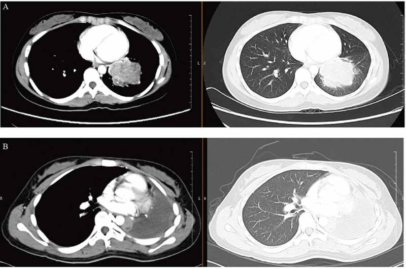
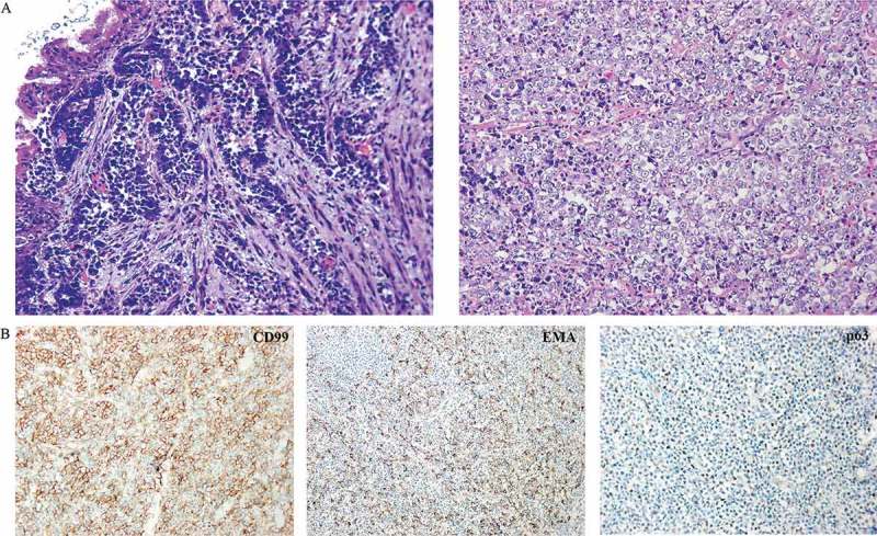
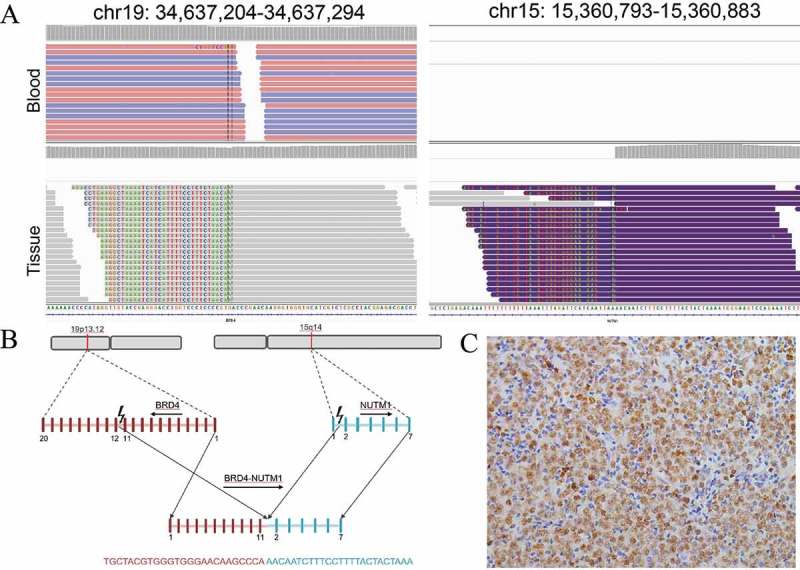
NUT carcinoma (NC) is an aggressive squamous tumor characterized by NUT gene rearrangement, and the most common fusion form is BRD4-NUT. However, NC diagnosis is difficult for its rareness and often being confused with a variety of poorly differentiated tumors. A 21-year-old Chinese woman was referred to our hospital for cough and intermittent fever. Chest computed tomography (CT) imaging revealed a left lobe hilar mass. Fiberoptic bronchoscopy results showed that tumor cells were poorly differentiated. In combination with immunohistochemistry staining, she was misdiagnosed with Ewing's sarcoma/primitive neuroectodermal tumor. Next-generation sequencing (NGS) revealing BRD4-NUT fusion, and NUT immunohistochemistry confirmed the diagnosis of NC. Subsequently, left pneumonectomy and lymph node dissection were performed, and the patient received pemetrexed and lobaplatin treatment. NGS technology played an important role in NC diagnosis in this case, and it may have clinical use for rare cancer diagnosis and guidance of potential targeted therapies.

Keywords: BRD4-NUT; NUT carcinoma; next-generation sequencing.


Figures

Figure 1.
Figure 1.
Disease status at diagnosis (a) and after left pneumonectomy and lymph node dissection (b) by CT scan (axial). CT, computed tomography.
Figure 2.
Figure 2.
Clinicopathologic characteristics of this case. (a) Pathology image of fiberoptic bronchoscopy deposits by hematoxylin and eosin stain showing round undifferentiated tumor cells. (b) Immunohistochemistry staining of tumor cells from a resected specimen that are positive for CD99, EMA and p63.
Figure 3.
Figure 3.
Detail presentation of BRD4-NUT fusion. (a) Screenshot of Integrative Genomics Viewer demonstrating uniquely mapped paired-end reads in the intronic regions of BRD4 (Intron 11) on chromosome 19 and NUT (Intron 1) on chromosome 15. For no hybrid capture probes were designed for NUT gene, tissue sample showed aberrant arrangement reads. (b) Schematic representation of the translocation responsible for the generation of the BRD4-NUT fusion gene. (c) Immunohistochemistry staining of tumor cells from resected specimen were positive for anti-NUT antibody.
Figure 4.
Figure 4.
Disease status after three weeks of chemotherapy by CT scan (axial). CT, computed tomography.

References

    1. French C. NUT midline carcinoma. Nature Rev Cancer. 2014;14:149–150. - PubMed
    1. French CA. Pathogenesis of NUT midline carcinoma. Annu Rev Pathol. 2012;7:247–265. doi:10.1146/annurev-pathol-011811-132438. - DOI - PubMed
    1. Balamuth NJ, Womer RB. Ewing’s sarcoma. Lancet Oncol. 2010;11:184–192. doi:10.1016/S1470-2045(09)70286-4. - DOI - PubMed
    1. Lessnick SL, Ladanyi M. Molecular pathogenesis of Ewing sarcoma: new therapeutic and transcriptional targets. Annu Rev Pathol. 2012;7:145–159. doi:10.1146/annurev-pathol-011110-130237. - DOI - PMC - PubMed
    1. Bishop JA, French CA, Ali SZ. Cytopathologic features of NUT midline carcinoma: A series of 26 specimens from 13 patients. Cancer Cytopathol. 2016;124:901–908. doi:10.1002/cncy.21761. - DOI - PubMed

MeSH terms