Review
. 2019 Jan;195(1):15-24.
doi: 10.1111/cei.13223. Epub 2018 Nov 13.

Enterovirus infection and type 1 diabetes: unraveling the crime scene

Affiliations
Review

Enterovirus infection and type 1 diabetes: unraveling the crime scene

T Rodriguez-Calvo. Clin Exp Immunol. 2019 Jan.

Abstract

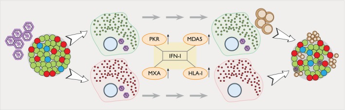
Enteroviruses (EV) have been historically associated to type 1 diabetes. Definitive proof for their implication in disease development is lacking, but growing evidence suggests that they could be involved in beta cell destruction either directly by killing beta cells or indirectly by creating an exacerbated inflammatory response in the islets, capable of attracting autoreactive T cells to the 'scene of the crime'. Epidemiological and serological studies have been associated with the appearance of islet autoimmunity and EV RNA has been detected in prospective studies. In addition, the EV capsid protein has been detected in the islets of recent-onset type 1 diabetic donors, suggesting the existence of a low-grade EV infection that could become persistent. Increasing evidence in the field shows that a 'viral signature' exists in type 1 diabetes and involves interferon responses that could be sustained during prolonged periods. These include the up-regulation of markers such as protein kinase R (PKR), melanoma differentiation-associated protein 5 (MDA5), retinoic acid inducible gene I (RIG-I), myxovirus resistance protein (MxA) and human leukocyte antigen-I (HLA-I) and the release of chemokines able to attract immune cells to the islets leading to insulitis. In this scenario, the hyperexpression of HLA-I molecules would promote antigen presentation to autoreactive T cells, favoring beta cell recognition and, ultimately, destruction. In this review, an overview is provided of the standing evidence that implicates EVs in beta cell 'murder', the time-line of events is investigated from EV entry in the cell to beta cell death and possible accomplices are highlighted that might be involved in beta cell demise.

Keywords: beta cell destruction; interferon response; type 1 diabetes; virus infection.


Figures

Figure 1
Figure 1
Schematic representation of the enterovirus genome. At the 5′ end the RNA contains an internal ribosomal entry site for cap‐dependent translation (IRES) and is covalently linked to the tyrosine‐3 residue of a small virus‐encoded peptide (VPg), which is used as a primer for RNA replication. The 3′ end has a polyadenylated tail [poly(A)] and contains an untranslated region (UTR). The genome encodes a single polyprotein that is cleaved into P1, P2 and P3. From the precursor P1, the capsid proteins VP0, VP3 and VP1 are generated. The non‐structural proteins 2A (protease) and 2ABC are formed from P2. Finally, P3 yields the production of 3AB and 3CD. The final processing steps lead to the production of VP4 and VP2 (capsid proteins), 2B (increases membrane permeability and inhibits cellular secretory pathways), 2C (vesicle formation), 3A (inhibits intracellular transport), 3B (VPg, primes RNA synthesis), 3C (protease) and 3D (polymerase).
Figure 2
Figure 2
Beta cells might fail to control enterovirus (EV) replication. EVs are able to replicate in both alpha and beta cells. Once the virus has entered the cells through its interaction with Coxsackie‐adenovirus receptor (CAR, viral RNA is sensed by protein kinase R (PKR) and melanoma differentiation‐associated protein 5 (MDA5), which induce the production of interferon (IFN)‐I and the creation of an anti‐viral state in the cell. This includes the up‐regulation of myxovirus resistance protein (MxA) (increasing IFN production) and islet human leukocyte antigen‐I (HLA‐I) expression. Due to a more efficient blocking of protein translation 8, alpha cells are able to stop viral production. In addition, IFN and other IFN‐related molecules are expressed at a higher level in alpha cells compared to beta cells, allowing them to rapidly eliminate the virus. Conversely, beta cells present a more sustained EV protein and IFN‐induced markers expression, reflecting a more chronic and non‐cytolytic infection 8. Additionally, beta cells can harbor persistent infections 11 and non‐cytopathic slow replicating viruses have been detected in the pancreas of persistently infected NOD mice 12, suggesting that beta cells might fail to control viral replication. Lastly, a sustained IFN‐stimulated HLA‐I hyperexpression could enhance beta cell antigen presentation, potentially leading to beta cell destruction by autoreactive CD8 T cells. Alpha cells are shown in red, beta cells in green, delta cells in blue, EVs in purple and CD8 T cells in brown.

References

    1. Tang JW, Holmes CW. Acute and chronic disease caused by enteroviruses. Virulence 2017;8:1062–5. - PMC - PubMed
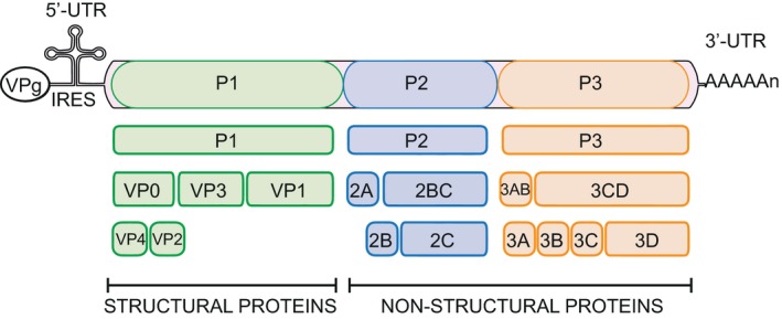
    1. Zell R. Picornaviridae – the ever‐growing virus family. Arch Virol 2018;163:299–317. - PubMed
    1. Racaniello VR. Picornaviridae: the viruses and their replicationIn Knipe DM, Howley PM, eds. Fields virology, vol. 1, 5th edn. Philadelphia, PA: Lippincott Williams & Wilkins; 2007, 795–838.
    1. van der Linden L, Wolthers KC, van Kuppeveld FJ. Replication and inhibitors of enteroviruses and parechoviruses. Viruses 2015;7:4529–62. - PMC - PubMed
    1. Shafren DR, Bates RC, Agrez MV, Herd RL, Burns GF, Barry RD. Coxsackieviruses B1, B3, and B5 use decay accelerating factor as a receptor for cell attachment. J Virol 1995;69:3873–7. - PMC - PubMed

Publication types