Review
. 2020 Jul;598(14):2957-2976.
doi: 10.1113/JP276962. Epub 2018 Nov 12.

Neurocardiac regulation: from cardiac mechanisms to novel therapeutic approaches

Affiliations
Review

Neurocardiac regulation: from cardiac mechanisms to novel therapeutic approaches

E N Bardsley et al. J Physiol. 2020 Jul.

Abstract

Cardiac sympathetic overactivity is a well-established contributor to the progression of neurogenic hypertension and heart failure, yet the underlying pathophysiology remains unclear. Recent studies have highlighted the importance of acutely regulated cyclic nucleotides and their effectors in the control of intracellular calcium and exocytosis. Emerging evidence now suggests that a significant component of sympathetic overactivity and enhanced transmission may arise from impaired cyclic nucleotide signalling, resulting from compromised phosphodiesterase activity, as well as alterations in receptor-coupled G-protein activation. In this review, we address some of the key cellular and molecular pathways that contribute to sympathetic overactivity in hypertension and discuss their potential for therapeutic targeting.

Keywords: Autonomic Nervous System; Cardiovascular Disease; Cyclic Nucleotide; Hypertension; Intracellular Calcium; Protein Kinase; Sympathetic Ganglion; Sympathetic Nervous System.


Figures

Figure 1
Figure 1. Sympathetic neurons are a powerful driver of myocyte function in cardiovascular disease
A, immunofluorescence depicting a co‐culture of sympathetic neurons and ventricular myocytes (reproduced from Larsen et al. 2016 b). Sympathetic neurons labelled with tyrosine hydroxylase (TH, green) densely innervate cultured cardiomyocytes labelled with sarcomeric α‐actinin (red). B, Wistar–Kyoto (WKY) or SHR sympathetic neurons were stimulated with nicotine (Nic) and the resulting myocyte cAMP was measured as a surrogate for sympathetic transmission, in mycoytes transduced with a cAMP Förster resonance energy transfer (FRET) sensor. FRET sensors were maximally stimulated (max) with an adenylyl cyclase (AC) activator forskolin (25 M) and a non‐specific phosphodiesterase (PDE) inhibitor 3‐isobutyl‐1‐methylxanthine (IBMX,100 M). In healthy co‐cultures (WKYn/WKYm), neuron‐evoked myocyte cAMP (17.05 ± 3.715, n = 29 cells) was significantly lower than cAMP measured in the diseased co‐culture myocytes (SHRn/SHRm; 44.02 ± 5.310, n = 36 cells; P < 0.0001). Cross‐cultures were established by plating diseased SHR neurons on top of healthy WKY myocytes (SHRn/WKYm) or healthy WKY neurons on top of diseased SHR myocytes (WKYn/SHRm). In the first cross‐culture (SHRn/WKYm), neuronal stimulation elevated myocyte cAMP (31.37 ± 5.194, n = 42 cells) to levels that were not significantly different from measured in the diseased (SHRn/SHRm) co‐cultures (P = 0.094), demonstrating that enhanced neuronal transmission elevates healthy‐myocyte cAMP to levels observed in disease. Moreover, in the second cross‐culture (WKYn/SHRm), stimulation of WKY neurons elevated SHR myocyte cAMP (15.67 ± 1.936, n = 24 cells) to levels that were not significantly different from that measured in healthy (WKYn/WKYm) co‐cultures (P = 0.76), demonstrating that healthy neurons attenuate the elevated myocyte cAMP response observed in SHR myocytes (modified from Larsen et al. 2016 b).
Figure 2
Figure 2. N‐type Ca2+ channel conductance is elevated in preSHR cardiac sympathetic neurons
Whole cell voltage clamp was performed on cardiac sympathetic stellate neurons to investigate whole cell Ca2+ currents. A, the current–voltage relationship. Access to the cell was obtained in normal Tyrode's solution containing the following (in mM): 135 NaCl, 4.5 KCl, 11 glucose, 20 HEPES, 1MgCl2, 2 CaCl2, pH 7.4. To identify the Ca2+ current, normal Tyrode solution was replaced with a Ca2+‐isolating solution using Ba2+ as the charge carrier, containing the following (in mM): 135 TEACl, 10 HEPES, 4.5 KCl, 1 MgCl2, 4 glucose, 1 NaHCO3, 2 BaCl2, pH 7.40, either in the presence or absence of ω‐conotoxin GVIA (1 μM), which selectively blocks N‐Type Ca2+ channels (IC50 = 0.15 nM) (Sato et al. 1993). Ba2+ was used as the charge carrier to avoid Ca2+‐dependent current inactivation (Imredy & Yue, 1994). The internal solution contained the following (in mM): 140 CsCl, 10 HEPES, 0.1 CaCl2, 1 MgCl2, 4 MgATP, 1 EGTA, pH 7.30. All solutions had osmolarities of 300 mOsm L−1. B, the whole cell Ca2+ current is larger in preSHR sympathetic nerves (127.5 ± 5.94 pA pF−1, n = 10) compared to WKY cells (−108.0 ± 6.80 pA pF−1, n = 10, P = 0.045) where peak current was recorded at −10 mV. ω‐Conotoxin GVIA (1 μM), significantly reduced the N‐type Ca2+ channel current to similar levels in both strains. A 75% reduction was observed in cells cultured from WKY stellate ganglia (−26.88 ± 1.7 pA pF−1, n = 6) and an 83% reduction was measured in neurons cultured from preSHR ganglia (−22.04 ± 1.60 pA pF−1, n = 5, ns) where peak current remained at −10 mV. Solid lines represent the mean of the WKY (black) and preSHR (red) control data. Dashed lines represent the mean of WKY (black) and preSHR (red) in the presence of ω‐Conotoxin GVIA. Data are represented as mean ± SEM. (A and B modified from Larsen et al. 2016 a).
Figure 3
Figure 3. Elevated Ca2+ conductance in preSHR stellate neurons is rescued with cGMP administration
AC, to ascertain whether cGMP signalling inhibits Ca2+ currents, whole cell voltage clamp was performed on sympathetic neurons young normotensive WKY (A) and young prehypertensive SHR (B) in the presence of a cGMP analogue, 8‐bromo‐cGMP (8b‐cGMP) (Larsen et al. 2016 a). Access to the cell was obtained in normal Tyrode solution containing the following (in mM): 135 NaCl, 4.5 KCl, 11 glucose, 20 HEPES, 1 MgCl2, 2 CaCl2, pH 7.4. To identify the Ca2+ current, the solution was replaced with a Ca2+‐isolating solution using Ba2+ as the charge carrier, containing the following (in mM): 135 TEACl, 10 Hepes, 4.5 KCl, 1 MgCl2, 4 glucose, 1 NaHCO3, 2 BaCl2, pH 7.40, in either the presence or the absence of 8b‐cGMP (100 μM). Ba2+ was used as the charge carrier to avoid Ca2+‐dependent current inactivation (Imredy & Yue, 1994). The internal solution contained the following (in mM): 140 CsCl, 10 Hepes, 0.1 CaCl2, 1 MgCl2, 4 MgATP, 1 EGTA, pH 7.30. All solutions had osmolarities of 300 mOsm L−1. 8b‐cGMP significantly reduced the elevated preSHR Ca2+ currents (−127.5 ± 5.94 pA pF−1, n = 10 to −105.2 ± 7.79 pA pF−1, n = 7) to levels that were no longer greater than WKY Ca2+ currents (−108.0 ± 6.80 pA pF−1, n = 10). Moreover, 8b‐cGMP had no significant effect on the WKY Ca2+ current, where peak currents were measured at −10 mV. Continuous lines represent the mean of the WKY (black) and preSHR (red) control data. Dashed lines represent the mean of WKY (black) and preSHR (red) in the presence of 8b‐cGMP. Data are represented as mean ± SEM. (AC are reproduced from Larsen et al. 2016 a). D, model diagram representing N‐type Ca2+ channel control by PKA and PKG, where PKA augments and PKG inhibits channel conductance. Pathways that are decreased (blue) or increased (pink) in disease are represented. AP, action potential; NA, noradrenaline; VGCC, voltage‐gated calcium channel; AC, adenylyl cyclase.
Figure 4
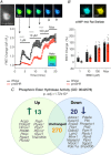
Figure 4. Phosphodiesterase (PDE) activity is impaired in preSHR neurons and has a genetic component
A and B, to investigate whether cytosolic PDE signalling is impaired in preSHR sympathetic neurons, a non‐specific PDE inhibitor, 3‐isobutyl‐1‐methylxanthine (IBMX; inhibits PDEs 1–7, 10–11), was administered to sympathetic stellate neurons (1–100 μM). The resulting intracellular cAMP was measured using real‐time Förster resonance energy transfer (FRET) in cells transduced with the adenovirus encoding the Epac‐SH187 biosensor (Klarenbeek et al. 2015). A, there was significantly greater IBMX‐stimulated cAMP in Wistar vs. preSHR neurons at all concentrations measured (two‐way repeated measures ANOVA; P < 0.05) supporting the evidence that there is a differential PDE profile in preSHR vs. control stellate neurons. At 100 μM IBMX, FRET responses were close to sensor saturation. B, peak FRET changes are depicted. Data are expressed as mean ± SEM. C, we investigated whether transcriptomic changes could be identified in SHR stellate ganglia with established hypertension. Using RNA sequencing, it was observed that the molecular function gene ontology (GO) group encoding ‘phosphoric ester hydrolase activity’ (GO:0042578) was significantly over‐represented in the SHR ganglia at 16 weeks. Thirty‐three genes were found to be differentially expressed and many of these mapped to regulators of PDE and kinase activity (figure reproduced from Bardsley et al. 2018 a).
Figure 5
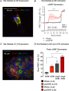
Figure 5. β‐AR signalling is elevated in preSHR neurons
A, we identified the presence of β‐adrenergic receptors (β‐ARs) on tyrosine hydroxylase (TH) positive cardiac sympathetic neurons. B, activation of presynaptic β‐ARs with isoprenaline (10 nM) led to a significantly larger cAMP generation in preSHR (56%; n = 12) vs. Wistar neurons (7%; n = 12; 2‐way ANOVA; P < 0.001), which was measured using real‐time cAMP in cells expressing the Epac‐SH187 biosensor (A and B are reproduced from Bardsley et al. 2018 b). C, we also identified the presence of AT1Rs on TH positive neurons. D, we investigated whether AT1R could elevate B1‐AR‐evoked cAMP. Dobutamine (DOB) alone elevated cAMP in preSHR neurons (reproduced from Bardsley et al. 2018 b). Moreover, AngII augments DOB‐evoked cAMP generation in Wistar neurons (n = 5, 6; P = 0.0073) and SHR neurons (n = 10, 8; P = 0.0005). We also measured a strain‐dependent effect following administration of DOB only (P = 0.0015) and in the presence of DOB with AngII (P = 0.0283). Data are represented as mean ± SEM.
Figure 6
Figure 6. Adrenaline is released from preSHR neurons
A, the catecholamine synthesis pathway, highlights the role of Phenylethanolamine‐N‐methyltransferase (PNMT) in the conversion from noradrenaline (NA) to Adrenaline (Adr). B and C, tyrosine hydroxylase (TH) and PNMT were measured in adult rat (B) and human (C) stellate ganglia (reproduced from Bardsley et al. 2018 b). D, using high pressure liquid chromatography with electrochemical detection (HPLC‐EC), we measured significantly higher total NA in Wistar (43.3 ± 2.173 pg; n = 8) compared with preSHR neurons (29.82 ± 6.366 pg; n = 4; P = 0.0294). In the same samples, we also measured a significantly greater total content of Adr in preSHR (14.14 ± 5.399 pg) compared with that measured in Wistar ganglia (3.937 ± 0.820 pg, P = 0.0019). E, electrical field stimulation of whole rat stellate ganglia led to the release of NA that was significantly higher in samples obtained from preSHR (4.32 ± 1.523 pg) vs. Wistar ganglia (1.477 ± 0.316 pg; P = 0.0396). The concentrations of neurally mediated Adr release were also significantly higher in preSHR (4.424 ± 1.391 pg, n = 4) compared with Wistar stellates (0.3201 ± 0.0325 pg; n = 8; P = 0.0028) (figure reproduced from Bardsley et al. 2018 b).

Similar articles

Cited by

References

    1. Abboud FM, Eckstein JW & Zimmerman BG (1964). Effect of dichloroisoproterenol on vascular responses to catecholamines. J Clin Invest 43, 316–322. - PMC - PubMed
    1. Adams MA, Bobik A & Korner PI (1989). Differential development of vascular and cardiac hypertrophy in genetic hypertension. Relation to sympathetic function. Hypertension 14, 191–202. - PubMed
    1. Ajijola OA, Vaseghi M, Mahajan A & Shivkumar K (2014). Bilateral cardiac sympathetic denervation: why, who and when? Expert Rev Cardiovasc Ther 10, 947–949. - PMC - PubMed
    1. Akaike T, Du N, Lu G, Minamisawa S, Wang Y & Ruan H (2017). A sarcoplasmic reticulum localized protein phosphatase regulates phospholamban phosphorylation and promotes ischemia reperfusion injury in the heart. JACC Basic Transl Sci 2, 160–180. - PMC - PubMed
    1. Apparsundaram S & Eikenburg DC (1995). Role of prejunctional beta adrenoceptors in rat cardiac sympathetic neurotransmission. J Pharmacol Exp Ther 272, 519–526. - PubMed

Publication types